العربية
Comprehensive Master Guide · Medically Reviewed

Carpal Compression Test: Your Guide to Accurate Wrist & Hand Diagnosis

CHAPTER 4 WRIST AND HAND ‌ ● A LIGAMENT/INSTABILITY TESTS 114 P iano key test 114 R adial collateral ligament stress test 115 Ulnar collateral ligament stress …

267 Detailed Chapters
33 min read
Updated: مارس 2026
Dr. Mohammed Hutaif
Medically Reviewed by
Prof. Dr. Mohammed Hutaif
Verified Content Expert Reviewed

Quick Medical Answer

In this comprehensive guide, we discuss everything you need to know about Carpal Compression Test: Your Guide to Accurate Wrist & Hand Diagnosis. A carpal compression test is a diagnostic maneuver used to assess for Carpal Tunnel Syndrome. It involves applying direct pressure over the median nerve at the carpal tunnel for up to 30 seconds. A positive carpal compression test reproduces symptoms like tingling, numbness, or pain in the median nerve distribution, indicating potential median nerve impingement.

Carpal Compression Test: Your Guide to Accurate Wrist & Hand Diagnosis

CHAPTER 4: WRIST AND HAND

A. LIGAMENT/INSTABILITY TESTS
* Piano key test
* Radial collateral ligament stress test
* Ulnar collateral ligament stress test
* Scaphoid shift test
* Lunotriquetral shear test
* Capitate apprehension test

B. TRIANGULAR FIBROCARTILAGE COMPLEX (TFCC) TESTS
* TFCC load test

C. THUMB TESTS
* Axial compression test
* Finkelstein’s test
* Ulnar collateral ligament laxity test

D. CARPAL TUNNEL TESTS
* Phalen’s test
* Tinel’s test
* Carpal compression test


A. LIGAMENT/INSTABILITY TESTS

Piano Key Test

Purpose
To detect the presence of instability at the inferior radio-ulnar joint (IRUJ).

Technique
* Patient position: Sitting with the forearm fully pronated and supported on a table.
* Clinician position: Sitting facing the patient, one hand stabilizes the patient’s hand in a neutral position while, approaching from the radial aspect, the index and middle fingers of the other hand are placed over the head of the ulna with the thumb providing some counterpressure under the base of the radius.
* Action: Downward pressure is applied by the fingers on the distal ulna, mimicking the action of pressing down a piano key.
* Positive test: Pain accompanied by excess movement and a loss of the normal ligamentous end-feel is noted.

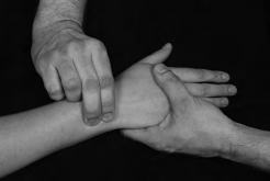
FIG 4.1: Piano key test.

Clinical context
Isolated involvement of the IRUJ is rare and is usually associated with more significant injury at the wrist (i.e. fracture or dislocation). Minor disruption may cause pain as the primary sign and this would be well localized with tenderness easily elicited by palpation. Pain at the end of passive pronation and supination would also be provocative. Where ligament disruption has occurred, excess movement will be accompanied by apprehension during certain activities, particularly in weight-bearing positions where its lack of stability is more seriously tested.

The IRUJ is often involved in inflammatory arthritis and in more severe cases, the synovitis afflicting the joint causes a destructive process resulting in pronounced loss of rotation, dorsal prominence and instability of the ulna, as well as localized swelling and loss of the normal function of the adjacent extensor carpi ulnaris, a condition known as caput ulnae syndrome (Brown & Neumann 2004). Given the associated joint stiffness, despite the instability at the IRUJ, the piano key test may not yield a positive result.

**Clinical tip:** The joint line is easily palpable on the dorsum of the lower forearm. The width of the wrist dorsally can be divided into three equal portions, making the joint line the marker between the most medial and middle sections. If the result of this test is ambiguous, further accessory testing of the IRUJ can be done to detect changes in range and end-feel.
**EXPERT OPINION (★★★):** Especially useful to determine level of instability – only a very unstable joint will behave like a true ‘piano key’.

Radial Collateral Ligament Stress Test

Aka: Wrist adduction test

Purpose
To stress the radial collateral ligament (RCL) and lateral capsule of the wrist in order to detect pain and/or laxity.

Technique
* Patient position: Sitting with the wrist supported on a table.
* Clinician position: One hand fixes the distal forearm by wrapping the fingers around the radius and ulna while the other grasps the hand, taking care not to involve the fingers or thumb.
* Action: With the forearm fixed, the distal hand takes the wrist into ulnar deviation (wrist adduction) where normal range is between 30° and 45°.
* Positive test: Pain is the most likely outcome but further evaluation would be needed if excessive range was noted suggesting significant disruption to the joint.

FIG 4.2: Radial collateral ligament stress test.

Clinical context
This test is most likely to identify an isolated sprain of the RCL where localized pain around the anatomical snuffbox would be the primary complaint. Depending on the severity of the trauma, a varying but equal degree of flexion/extension restriction may also be present at the wrist as the ligament blends with, and reinforces, the lateral joint capsule, thereby causing capsular limitation.

As the history is usually traumatic, careful screening is necessary (including X-ray evaluation in most cases) to eliminate fracture of the carpus—the scaphoid being the usual culprit. With pain reported in this area, osteoarthritis of the trapeziofirstmetacarpal joint (basal joint of the thumb; see axial compression test) and de Quervain’s tenosynovitis (see Finkelstein’s test) are other possible diagnoses to consider.

**Clinical tip:** The RCL can be found by identifying the radial styloid at the base of the anatomical snuffbox and moving the finger slightly distally with the wrist in a degree of radial deviation. The ligament and capsule are relaxed in this position. Keeping the finger in place, the RCL tautens (if intact) and becomes easily palpable as the wrist is passively taken into ulnar deviation. Make sure the patient’s thumb is not involved in this movement in order to avoid stress being placed on the thumb abductor and extensor tendons which may result in a false positive finding.
**EXPERT OPINION (★):** Usually included as part of radiocarpal assessment without specifically testing the ligaments as they are rarely injured in isolation.

Ulnar Collateral Ligament Stress Test

Aka: Wrist abduction test

Purpose
To stress the ulnar collateral ligament (UCL) and medial capsule of the wrist in order to detect pain and/or laxity.

Technique
* Patient position: Sitting with the wrist supported on a table.
* Clinician position: One hand fixes the distal forearm by wrapping fingers around the radius and ulna while the other grasps the hand, taking care not to involve the fingers or thumb.
* Action: With the forearm fixed, the distal hand takes the wrist into radial deviation (wrist abduction) where normal range is around 15°.
* Positive test: Pain is the most likely outcome but further evaluation would be needed if excessive range was noted, suggesting significant disruption to the joint.

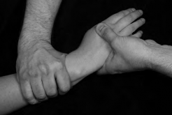
FIG 4.3: Ulnar collateral ligament stress test.

Clinical context
The patient reports localized pain located at the inner, medial aspect of the wrist joint. If the UCL stress test is the only positive test, confirmation of the lesion can be made by palpation—tenderness is usually identifiable at its origin on the ulnar styloid. Isolated injury to the UCL is rare and, because of its anatomical connection, is more commonly associated with injury to the triangular fibrocartilaginous complex (see TFCC test). Medial wrist pain may also emanate from the inferior radio-ulnar joint (see piano key test) and the extensor carpi ulnaris tendon.

**Clinical tip:** The ligament is most easily identified by finding the styloid with the wrist in ulnar deviation and then passively moving the wrist radially until the ligament can be felt tautening under the finger.
**EXPERT OPINION (★):** Usually included as part of radiocarpal assessment without specifically testing the ligaments as they are rarely injured in isolation.

Scaphoid Shift Test

Aka: Watson test

Purpose
To establish the presence of abnormal movement of the scaphoid and lunate bones indicating instability or subluxation.

Technique
* Patient position: The patient is seated with the flexed elbow resting on a table, the forearm vertically positioned and fully pronated so that the patient’s palm faces the clinician.
* Clinician position: Seated facing the patient, the examiner places the thumb in the palm and wraps the fingers around the metacarpals on the dorsum of the hand. The thumb of the second hand is placed over the tubercle of the scaphoid and counterpressure applied with the other fingers over the dorsum of the lower radius.
* Action: Firm pressure is applied on the scaphoid while the hand is taken initially into ulnar deviation and slight extension to offload the scapholunate articulation. Maintaining the pressure on the scaphoid, the wrist is then taken slowly into radial deviation and slight flexion creating a subluxation force that stresses the articulation and exposes instability if present.
* Positive test: Excessive movement of the scaphoid in relation to the lunate is detected along with some pain and/or apprehension. If subluxation occurs the proximal pole of the scaphoid shifts dorsally over the dorsal rim of the radius. Removing the pressure from the palmar aspect of the scaphoid and returning the wrist into some ulnar deviation and extension will cause the scaphoid to shift back in a palmar direction to its reduced, normal position.

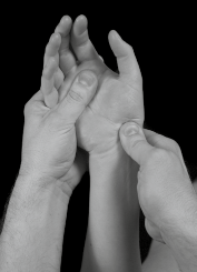
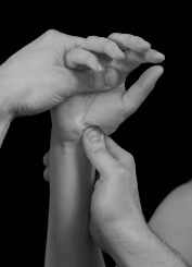
FIG 4.4: Scaphoid shift test: unloaded start position (A) and stress applied to the scapholunate joint (B).

Clinical context
Although comparatively rare, the instability that results from disruption of the scapholunate articulation is the most common carpal instability. A complete tear of the scapholunate ligaments, usually resulting from a severe hyperextension injury, will lead to significant scapholunate dissociation and disruption to the normal motion of the proximal carpal bones during wrist movement. Because of the loss of connection between the scaphoid and lunate, the scaphoid rotates into a degree of flexion leaving the lunate and triquetral free to rotate into extension which causes pain, an inability to weight-bear on the wrist and an overall loss of function (Placzek & Boyce 2006).

Chronic instability of the joint also strongly predisposes to a recognized sequence of osteoarthritis involving the unstable capito-lunate and capitoscaphoid articulations, a sequence known as scapholunate advance collapse (SLAC) (Miller & Schweitzer 2005).

The normal extent of separation between the scaphoid and lunate bones should be less than 2 mm (Gross et al 2002) but a diastasis of more than 3 mm is pathognomonic of scapholunate dissociation and should be detectable on plain X-rays (McRae 1990). The evident space between the bones is known as the Terry Thomas sign , named after the comedian who famously had a gap between his two front teeth.

Using a cadaver model where the wrist was progressively loaded in extension and ulnar deviation, a sequential, four-staged pattern of injury was noted; scapholunate diastasis, dorsal subluxation of the capitate, disruption of the lunotriquetral ligament and complete dislocation of the scapholunate articulation (Brown & Neumann 2004).

**Clinical tip:** It is not always necessary to reproduce subluxation with this test as it is usually painful for the patient and they are likely to indicate apprehension as the forces are gradually applied. The presence of pain and apprehension are good indicators of instability and, as an alternative, detecting increased excursion without causing subluxation can be achieved by applying an anterior/posterior glide to the scaphoid in a neutral position and comparing with the opposite side.
**EXPERT OPINION (★★★):** A tricky test to perform, as well as interpret, but very useful once perfected. Care should be taken when interpreting the findings as up to 30% of healthy wrists can give a false positive result due to general ligamentous laxity at the wrist. It is thought that repeating this dynamically (see variations), particularly when the patient makes a fist, reduces this tendency (Weiss & Finkelstein 2005).

Variations
The scaphoid stress test is a simple modification where the patient actively moves the wrist from ulnar to radial deviation while the scaphoid is stabilized by the examiner in the same way. Apprehension and/or a painful clunk during the movement demonstrates dynamic subluxation.


Lunotriquetral Shear Test

Aka: Ballottement test, Reagan’s test, Shuck test

Purpose
To establish the presence of abnormal movement of the lunate and triquetral bones indicating instability or subluxation.

Technique
* Patient position: The patient is seated with the forearm supinated and comfortably supported on a table.
* Clinician position: Seated facing the patient, the clinician, using a pincer grip, stabilizes the lunate by placing a thumb on its palmar surface and fixes the dorsal surface with the index finger. The other hand grasps the combined bony mass of the pisiform and triquetral using the same pincer grip.
* Action: With the lunate stabilized, the examiner moves the triquetral/pisiform in an anteroposterior direction. The test can be reversed by stabilizing the triquetral/pisiform and moving the lunate in the same plane.
* Positive test: Excessive mobility of the triquetral in relation to the lunate when compared to the unaffected side is detected along with pain. Crepitus on movement is also sometimes noted.

FIG 4.5: Lunotriquetral shear test.

Clinical context
Differing descriptions for this test can be found in the literature and this has led to the same test being assigned several names. Injury to the lunotriquetral articulation is uncommonly encountered requiring impact in extension and radial deviation (Brown & Neumann 2004). A strain to the lunotriquetral ligament may be evident with localized tenderness and a positive test, but if the trauma has been sufficiently significant to cause disruption of the articulation, normal kinematics will be lost.

The combined unit of the scaphoid and lunate rotate into flexion leaving the untethered triquetral to drift into extension. Pain, apprehension, a reluctance to move and weakness will all be reported by the patient, with a more significant injury likely to predispose to osteoarthritis (Placzek & Boyce 2006). Lunotriquetral ligament injury is often associated with triangular fibrocartilage complex (TFCC) tears and MRI can therefore be helpful in making the distinction (Miller & Schweitzer 2005).

**Clinical tip:** An isolated injury to the lunotriquetral ligament does not usually produce a static diastasis between the lunate and triquetral so there may be dynamic and functional instability present but a normal plain X-ray reported.
**EXPERT OPINION (★★):** It is important to establish what is normal excursion on the unaffected side as differences in range can be subtle.

Related tests
Murphy’s sign may also indicate the presence of lunate dislocation. The patient is asked to make a fist and the relationship between the heads of the 2nd, 3rd and 4th metacarpals is examined. Normally the head of the 3rd metacarpal extends more distally but where the lunate has dislocated, it remains in line with the heads of the other two metacarpals.


Capitate Apprehension Test

Aka: Capitate displacement test

Purpose
To determine the presence of capitate instability.

Technique
* Patient position: The patient sits with the forearm supinated and supported on a table.
* Clinician position: Facing the patient, one of the examiner’s thumbs is placed over the palmar aspect of the capitate bone reinforced with the thumb of the other hand. The fingers of both hands are then wrapped around the dorsum of the patient’s hand ensuring the wrist is supported in a neutral position and taking care with the handhold not to involve the patient’s thumb.
* Action: The examiner pushes the capitate posteriorly with both thumbs, ensuring the hand is stabilized by the fingers placed around the dorsum of the wrist.
* Positive test: Pain and/or apprehension is reproduced as the posterior pressure is applied and this is sometimes accompanied by a click.

FIG 4.6: Capitate apprehension test.

Clinical context
Capitate subluxation is most commonly associated with instability at the scapholunate articulation, usually resulting from a fall on an outstretched hand. Laxity or rupture of the scapholunate ligaments creates a static or dynamic diastasis that leaves the proximal capitate vulnerable to dorsal subluxation, either at the time of the injury or subsequently. In the presence of laxity, the capitate may sublux and reduce recurrently. When subluxed, the capitate can become more prominent and this is best observed on the dorsum of the hand with the wrist in a flexed position. Passive wrist extension will also be painfully blocked particularly when weight-bearing (Atkins et al 2010).

**Clinical tip:** The capitate is located at the base of the 3rd metacarpal and can more easily be palpated on the dorsum of the wrist. There is normally a palpable ‘dip’ over the capitate in the neutral position although this is lost if subluxation has occurred.
**EXPERT OPINION (★★):** Only used when a generalized mid-carpal instability is suspected with a patient complaining of episodic subluxation and pain.

Related tests
The midcarpal pivot shift test aims to identify instability as a result of traumatic attenuation of the scapholunate or lunotriquetral ligaments as midcarpal instability is often associated with concurrent injury to these articulations. The patient sits with the elbow at 90° with the supinated forearm supported on a table. The examiner stabilizes the distal forearm with one hand and, supporting the patient’s hand in a neutral position with the other, takes the wrist from a fully radially deviated position into full ulnar deviation. Instead of a normal smooth movement, it is irregular and accompanied by a painful clunk which indicates a positive finding.

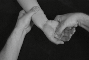
FIG 4.7: Midcarpal pivot shift. Start position (A). End position stressing the scapholunate and lunotriquetral articulations (B).

Lichtman’s test is a variation of the midcarpal pivot shift test where axial compression is added to the movement. Pain and/or dorsal movement of the capitate are considered to be positive findings.

**EXPERT OPINION (★) for Lichtman’s Test:** This test can be hard to do well and interpret the findings accurately.

B. TRIANGULAR FIBROCARTILAGE COMPLEX (TFCC) TESTS

TFCC Load Test

Aka: Ulnar meniscal grind test

Purpose
To reproduce pain and/or apprehension indicating a tear or degeneration of the TFCC.

Technique
* Patient position: Sitting or standing.
* Clinician position: Facing the patient, the examiner stabilizes the patient’s forearm with one hand and, as if shaking hands, places their other hand in the palm where it is held firmly.
* Action: Axial compression is then applied through the patient’s hand while ulnar deviation is added. This part of the manoeuvre has been described as the ulnar impaction test . This may be enough to reproduce localized pain at the base of the ulna negating the need for further loading. If asymptomatic, stress on the TFCC is increased by ‘scooping’ the hand from flexion to extension while maintaining the ulnar deviation and compression.
* Positive test: Localized pain at the ulnar side of the wrist joint is sometimes accompanied by apprehension and/or a click or crepitus on movement.

FIG 4.8: Triangular fibrocartilage complex (TFCC) test.

Clinical context
The TFCC, sometimes referred to as the ulnar articular disc, is a homogeneous structure comprising of the dorsal and palmar radioulnar ligaments, a meniscus, the ulnar collateral ligament and the sheath of the extensor carpi ulnaris (Palmer & Werner 1981). It acts as the primary soft tissue stabilizer of the distal radioulnar joint and takes 20% of the compressive load across the wrist. It is thickest (approx. 5 mm) at its ulnar insertion but thinner (2 mm) and more vulnerable to injury nearer to its radial origin which, unsurprisingly, is the most common site of TFCC tears (Miller & Schweitzer 2005).

The central 80% of the TFCC is avascular and has little potential for repair although its periphery and the dorsal and palmar ligaments are well vascularized, opening the possibility for healing in this zone (Bulstrode et al 2002).

Traumatic lesions (class one) usually involve a compressive force with rotation and/or ulnar deviation. The injury can be masked by associated fractures of the radius and/or ulna. Degenerative/over-use lesions (class two) are found increasingly over the age of 30 and in some cases may progress from simple TFCC ‘wear’ to accompanying lunotriquetral ligament disruption and ulnocarpal arthritis (Bulstrode et al 2002, Miller & Schweitzer 2005).

**Clinical tip:** Clinical features of TFCC lesions include wrist pain accentuated by movement, particularly pronation and ulnar deviation as well as loading of the clenched fist. These findings are usually accompanied by tenderness and crepitus over the TFCC area (Bulstrode et al 2002).
**EXPERT OPINION (★★★):** A very useful test but it can produce false positives and should therefore be taken in conjunction with all other findings.

Related tests
The supination lift test requires the patient to sit facing an examination couch with the elbows flexed to 90° and the forearms fully supinated. The couch is positioned at a height that allows the palms of both hands to comfortably make contact with its under-surface. The patient is then asked to attempt to lift the couch up. Localized pain at the base of the ulnar and a reluctance to apply full force are both indicative of a TFCC tear.


C. THUMB TESTS

Axial Compression Test

Aka: Axial grind test, Thumb grind test, Basal joint grind test

Purpose
To detect osteoarthritis (OA) of the trapeziofirstmetacarpal joint (basal joint of the thumb).

Technique
* Patient position: The hand rests in a mid-pronated position on a table.
* Clinician position and action: The compression test is performed by stabilizing the radial aspect of the wrist with one hand and gripping the first metacarpal shaft with the fingers and thumb of the other. An axial load is applied downwards along the shaft while the metacarpal is gently moved against the trapezium. This has been described as a ‘grind’ technique as the articular surfaces are moved together under compression.
* Positive test: A sudden, sharp pain is usually elicited as compression is applied. Occasionally crepitus may also be noted.

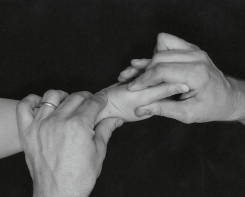
FIG 4.9: Axial compression test of the trapeziofirstmetacarpal joint. The arrow indicates the direction of axial compression.

Clinical context
OA of the trapeziofirstmetacarpal joint is the most common site of degenerative joint disease in the hand (Brown & Neumann 2004, Ghavami & Oishi 2006, McRae 1990). The pain is localized around the base of the thumb and typically described as ‘piercing’ and exacerbated by twisting and gripping motions making functional tasks difficult. In the early stages, before changes become detectable on X-ray, the irritated joint capsule may begin to develop capsular restriction causing loss of extension (Atkins et al 2010).

At this stage, in the absence of articular changes, the axial compression test is unlikely to be positive but as the condition worsens, laxity of the supporting ligaments and a reduction of bony constraints leads to increasing joint stress and progressive degenerative disease. Once established, the sensitivity of the test increases but diagnosis is ultimately confirmed by radiology.

Trapeziofirstmetacarpal Osteoarthritis Staging (Eaton & Glickel 1987)
* Stage I: Mild joint narrowing or subchondral sclerosis noted with small effusion. No laxity, subluxation or osteophyte formation.
* Stage II: Possible osteophyte formation at the ulnar side of the distal trapezial articular surface. Mild to moderate subluxation might appear.
* Stage III: Further joint space narrowing with cystic changes, bone sclerosis and prominent osteophytes. The first metacarpal is moderately subluxed radially and dorsally. Passive reduction might not be possible.
* Stage IV: Scaphotrapezial joint also involved with evidence of destruction, with the trapeziofirstmetacarpal joint immobile and usually largely pain-free.

**Clinical tip:** Care needs to be taken when assessing this joint as axial loading can elicit severe twinges of pain, particularly when the condition presents acutely.
**EXPERT OPINION (★★★):** Used regularly as part of the routine clinical examination – especially if X-rays are not available to grade the stage of osteoarthritis.

Finkelstein’s Test

Purpose
To detect pain and limitation caused by inflammation between the tendons of abductor pollicis longus (APL) and extensor pollicis brevis (EPB) and their shared synovial sheath at the distal end of the radius (de Quervain’s tenosynovitis).

Technique
* Patient position: With the forearm positioned in pronation, the patient is asked to flex the thumb and close their fingers over it.
* Clinician position and action: The lower forearm is fixed with one hand and the patient’s hand taken into ulnar deviation passively with the other.
* Positive test: As the wrist is taken towards ulnar deviation, significant pain is reproduced over the radial aspect of the wrist.

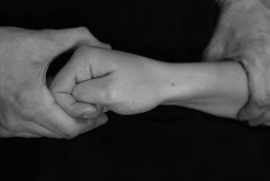
FIG 4.10: Finkelstein’s test.

Clinical context
De Quervain’s tenosynovitis involves the two tendons in the first dorsal synovial compartment of the wrist. In the chronic stage where adhesions have developed and the sheath has become thickened, the condition is known as de Quervain’s stenosing tenosynovitis (Atkins et al 2010, Placzek & Boyce 2006). The patient reports localized pain over the dorsum of the distal radius and this is often accompanied by swelling and crepitus on movement. Isometric resisted testing of the affected tendons is painful but repeated active thumb extension more so. The history is usually one of overuse or unaccustomed activity.

The incidence is reported to be considerably higher in the female population, with age also being a significant risk factor and prevalence increasing in the 40+ age group (Wolf et al 2009). The relative excursions of the APL and EPB tendons during Finkelstein’s testing suggest that a positive Finkelstein’s test may result more from EPB pathology than the APL (Kutsumi et al 2005).

**Clinical tip:** Even in the asymptomatic hand this test can be uncomfortable and care should be taken to ensure that the pain is not excessively provoked by an over-vigorous technique, particularly when acutely painful. De Quervain’s tenosynovitis should not be confused with intersection syndrome which involves the same two tendons as they cross over the wrist extensor muscles in the lower forearm about 5 cm proximal from their sheathed extent (Atkins et al 2010) and, because of this, Finkelstein’s test should not be significantly provocative.
**EXPERT OPINION (★★):** This can be approached with too much vigour. A subtle test if done well.

Ulnar Collateral Ligament Laxity Test

Purpose
To detect pain and/or laxity of the ulnar collateral ligament (UCL) of the first metacarpophalangeal (MCP) joint.

Technique
* Patient position: Seated with the hand supported on a table in a mid-pronated position.
* Clinician position: The examiner stabilizes the distal end of the first metacarpal with the thumb and index finger. The same pincer grip is used with the other hand, placing the fingers over the radial and ulnar aspects of the proximal phalanx.
* Action: Fixing the metacarpal, a valgus stress is applied to the joint (which in this position involves the thumb being moved towards the patient).
* Positive test: Pain and/or laxity reproduced at the base of the thumb.

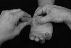
FIG 4.11: Valgus stress on the ulnar collateral ligament of the first MCP joint.

Clinical context
Gamekeeper’s thumb (Campbell 1955) is caused by a chronic insufficiency of the UCL at the first MCP joint leading to pain and weakness of the ‘pinch grasp’. Injury to the UCL also presents acutely, most commonly among the skiing population (skier’s thumb), as a result of the ski-pole abruptly stopping in the snow during a fall and the static handle forcing a sudden valgus or abduction stress to the thumb (Davidson & Laliotis 1996, Van Dommelen & Zvirbulis 1989).

When both the ulnar and accessory collateral ligaments are ruptured, a Stener lesion may develop, where the aponeurosis of the adjacent adductor brevis muscle becomes interposed between the ruptured ligament and the phalanx. The ligament retracts, no longer able to make contact with its insertion, and fails to heal (Stener 1962). As an important stabilizer of the thumb the loss of the UCL hampers function considerably and surgical repair is often necessary (Miller & Schweitzer 2005).

**Clinical tip:** With the joint in an extended position, valgus testing primarily determines the competence of the accessory collateral ligament which is taut in this position. Laxity of between 15° and 30° would indicate rupture or partial rupture of the ligament and arouse suspicion of a gamekeeper’s fracture where a portion of the proximal phalanx at the UCL insertion becomes avulsed. A displaced fracture should be eliminated with X-ray before the clinician repeats the valgus test in 30° of flexion, as this position preferentially stresses the UCL and could disturb the fracture site. If gamekeeper’s fracture has been ruled out, an MRI or MR arthrography would be necessary to diagnose a Stener lesion (Harper et al 1996, Spaeth et al 1993).
**EXPERT OPINION (★★★):** In acute injuries this test can determine whether surgical intervention is required. Most surgeons will have a point at which they will repair the ligament (usually around 30–40° of laxity). However, it is a matter of weighing up a number of factors: age, occupation, patient choice, time elapsed since trauma, anaesthetic risk, etc., as this may alter the threshold for surgical repair.

D. CARPAL TUNNEL TESTS

Phalen’s Test

Purpose
To increase pressure on the median nerve as it passes through the carpal tunnel in order to aid diagnosis of carpal tunnel syndrome (CTS).

Technique
* Patient position: Seated with the hand resting on a table in mid-pronation.
* Clinician position: Seated facing the patient. The affected hand is taken into full wrist flexion. Alternatively, the patient is asked to flex both wrists and oppose the dorsum of the hands so that the flexion is maintained bilaterally.
* Action: In either position, the wrist flexion is maintained for a minute.
* Positive test: Paraesthesiae is reproduced in the cutaneous distribution of the median nerve (the palmar aspect of the thumb, index and middle fingers and the lateral half of the ring finger) as a result of the sustained narrowing at the carpal tunnel during flexion of the wrist. If severe, pain may also be reproduced.

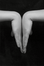
FIG 4.12: Phalen’s test (A) and alternative position (B).

Clinical context
Studies of CTS have generated a great deal of controversy and no universally accepted diagnostic criteria exist (Placzek & Boyce 2006). Phalen’s test was always widely considered to be the most sensitive physical test, but over recent years a number of studies have challenged this and attempted to demonstrate enhanced sensitivity with related or modified tests.

The absence of an agreed ‘gold standard’ of diagnosis requires the clinician to combine a number of clinical findings which can be used to predict the probability of CTS. The ‘CTS 6’ is a validated clinical diagnostic aid which assesses six factors (Graham 2008):
1. Numbness exclusively or predominantly in the median nerve distribution of the hand.
2. Nocturnal numbness.
3. Thenar atrophy or weakness (≤grade 4 on examination).
4. Positive Phalen’s test.
5. Loss of two-point discrimination (failure to distinguish two points 5 mm apart).
6. Positive Tinel’s test.

Table 4.1: Phalen’s Test Validity

Author and year LR + LR - Target condition
Golding et al 1986 0.71 1.05 CTS
Gerr & Letz 1998 1.3 0.8 CTS
Williams et al 1992 88 (★★★) 0.12 (★★) CTS
Tetro et al 1998 3.6 (★) 0.5 (★) CTS
**Clinical tip:** Differential diagnosis of hand paraesthesiae/pain should include cervical myelopathy, radiculopathy, adverse neurodynamics involving the lower cervical nerve roots, thoracic outlet syndrome and ulnar nerve entrapment syndromes at the elbow and wrist. Flattening of the thenar eminence muscles may be evident in more chronic cases.
**EXPERT OPINION (★★★):** This is the preferential test for CTS but the clinician should always be aware of the potential of a more proximal source of symptoms requiring further examination of the cervical spine and brachial plexus.

Additional tests
The prayer test can also be used to detect CTS. The affected hand is taken into full wrist extension (or together the palms of the hands are opposed and lowered until full extension at both wrists is achieved) and held there for a minute. A ‘decompression phenomenon’ is sometimes observed when pressure on the nerve trunk is removed.

FIG 4.13: Prayer test.

The tourniquet test attempts to exacerbate median neuropathy by inducing temporary ischaemia in the hand. The hand elevation test (Ahn 2001) simply involves elevating the hand for up to a minute to reproduce symptoms. The three jaw chuck test involves opposing the fingers with the thumb and then flexing the wrist maximally.

FIG 4.14: Three jaw chuck test.


Tinel’s Test

Aka: Median nerve percussion test

Purpose
To elicit paraesthesiae and/or pain in the median nerve distribution of the hand in order to aid diagnosis of carpal tunnel syndrome (CTS).

Technique
* Patient position: Seated with the hand resting on a table with the forearm fully supinated.
* Clinician position: Sitting facing the patient, the affected hand is held in a neutral position.
* Action: The mid-point of the carpal tunnel is identified and ‘tapped’ with a finger or a percussion hammer.
* Positive test: Temporary paraethesiae or pain in the cutaneous distribution of the median nerve in the hand is reported.

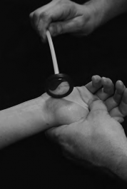
FIG 4.15: Tinel’s test at the carpal tunnel.

Clinical context
Tinel’s test is used in other compressive neuropathies, although it is best known for the detection of CTS. Wide variations in both sensitivity and specificity of the test have been reported. No physical test has been shown to be definitive although a combination of tests, including Tinel’s, was shown to yield reasonable detection rates (Graham 2008).

Table 4.2: Tinel’s Test Validity

Author and year LR + LR - Target condition
DeKrom et al 1990 0.6 1.3 CTS
Golding et al 1986 1.3 0.9 CTS
Gerr & Letz 1998 0.67 1.1 CTS
Tetro et al 1998 8.2 (★★) 0.3 (★) CTS
Williams et al 1992 67 (★★★) 0.3 (★) CTS
**Clinical tip:** Tinel’s test can also be used to detect the extent of regeneration of the sensory fibres of the median nerve where the most distal point of abnormal sensation represents the distal extent of the regeneration. The clinician commences tapping at the tip of the palmar surface of the index finger and moves proximally towards the mid-point of the carpal tunnel.
**EXPERT OPINION (★★):** Used mainly to determine continued potential for nerve recovery post op/trauma rather than actual diagnosis of the condition.

Related tests
Although less common, ulnar tunnel syndrome results from compression of the ulnar nerve as it passes through Guyon’s canal. It is usually caused by sustained compression (e.g. positioning the ulnar border of the hand on the handlebars when cycling with the wrist in extension and radial deviation). Tinel’s test can be repeated, tapping distally from the pisiform.

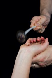
FIG 4.16: Tinel’s test at the ulnar tunnel.


Carpal Compression Test

Aka: Pressure provocative test, Manual carpal compression test, Durkan’s compression test

Purpose
To increase pressure on the median nerve as it passes through the carpal tunnel in order to aid diagnosis of carpal tunnel syndrome (CTS).

Technique
* Patient position: Seated with the forearm supinated with the hand rested on a table.
* Clinician position and action: Sitting facing the patient, the examiner places one thumb, superimposed with the other, over the mid-point of the flexor retinaculum and presses firmly downwards for up to a minute while maintaining counterpressure with the fingers on the dorsum of the hand.
* Positive test: Paraesthesiae are reproduced in the cutaneous distribution of the median nerve and if the condition is severe, pain may also be evoked.

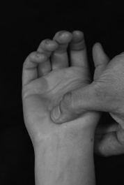
FIG 4.17: Carpal compression test.

Clinical context
The carpal compression test has been shown to be more sensitive than both Phalen’s and Tinel’s tests, with symptoms often reproducible within several seconds, suggesting that it is a simple, fast and valuable provocative test for CTS (González et al 1997). In an attempt to standardize the amount of pressure required to consistently reproduce the symptoms, the use of an instrumented carpal compression device was tested and shown to deliver a similar degree of sensitivity and specificity (Durkan 1994).

Table 4.3: Carpal Compression Test Validity

Author and year LR + LR - Target condition
Gonzalez et al 1997 7.9 (★★) 0.15 (★★) CTS
Durkan 1994 22.3 (★★★) 0.11 (★★) CTS
Tetro et al 1998 10.7 (★★★) 0.27 (★) CTS
Williams et al 1992 33 (★★★) 0.01 (★★★) CTS
**Clinical tip:** This test is also usefully employed where there is limited range of flexion or pain at the wrist which prevents an effective Phalen’s test from being performed. Accurate localization of the median nerve in the carpal tunnel is necessary for effective testing. The distal wrist crease is level with the proximal border of the flexor retinaculum so pressure needs to be distal to this line and centrally.

Variations
The test can be further modified by either adding passive wrist extension (while asking the patient to grip) or passive wrist flexion (Fertl et al 1998, Tetro et al 1998). The latter has been found to have an optimal cut-off time of just 20 seconds.

Table 4.4: Carpal Compression Test with Flexion/Extension

Author and year LR + LR - Target condition
Tetro et al 1998 82 (★★★) 0.18 (★★) CTS

Scientific References

  • Ahn, D.S., 2001. Hand elevation: a new test for carpal tunnel syndrome. Ann. Plast. Surg. 46 (2), 120–124.
  • Atkins, E., Kerr, E., Goodlad, J., 2010. A Practical Approach to Orthopaedic Medicine , third ed. Churchill Livingstone, Edinburgh.
  • Brown, D.E., Neumann, R.D., 2004. Orthopaedic Secrets , third ed. Hanley & Belfus, Philadelphia.
  • Bulstrode, C., Buckwalter, J., Carr, A., et al., 2002. Oxford Textbook of Orthopaedics and Trauma . Oxford University Press, Oxford.
  • Campbell, C.S., 1955. Gamekeeper’s thumb. J. Bone Joint Surg. Br. 37 (1), 148–149.
  • Davidson, T.M., Laliotis, A.T., 1996. Snowboarding injuries, a four-year study with comparison with alpine ski injuries. West. J. Med. 164 (3), 231–237.
  • DeKrom, M.C., Knipschild, P.G., Kester, A.D., 1990. Efficacy of provocative tests for diagnosis of carpal tunnel syndrome. Lancet 335 (8686), 393–395.
  • Durkan, J.A., 1994. The carpal-compression test: an instrumented device for diagnosing carpal tunnel syndrome. Orthop. Rev. 23 (6), 522–525.
  • Eaton, R.G., Glickel, S.Z., 1987. Trapeziometacarpal osteoarthritis: staging as a rationale for treatment. Hand Clin. 3 (4), 455–471.
  • Fertl, E., Wober, C., Zeitlhofer, J., 1998. The serial use of two provocative tests in the clinical diagnosis of carpal tunnel syndrome. Acta Neurol. Scand. 98 (5), 328–332.
  • Gerr, F., Letz, R., 1998. The sensitivity and specificity of tests for carpal tunnel syndrome vary with the comparison subjects. J. Hand Surg. Br. 23 (2), 151–155.
  • Ghavami, A., Oishi, S.N., 2006. Thumb trapeziometacarpal arthritis: treatment with ligament reconstruction tendon interposition arthroplasty. Plast. Reconstr. Surg. 117 (6), 116e–128e.
  • Golding, D.N., Rose, D.M., Selvarajah, K., 1986. Clinical tests for carpal tunnel syndrome: an evaluation. Br. J. Rheumatol. 25 (4), 388–390.
  • González del Pino, J., Delgado-Martínez, A.D., González González, I., et al., 1997. Value of the carpal compression test in the diagnosis of carpal tunnel syndrome. J. Hand Surg. Br. 22 (1), 38–41.
  • Graham, B., 2008. The value added by electrodiagnostic testing in the diagnosis of carpal tunnel syndrome. J. Bone Joint Surg. Am. 90 (12), 2587–2593.
  • Gross, J., Fetto, J., Rosen, E., 2002. Musculoskeletal Examination , second ed. Wiley-Blackwell, Chichester.
  • Harper, M.T., Chandnani, V.P., Spaeth, J., et al., 1996. Gamekeeper thumb: diagnosis of ulnar collateral ligament injury using magnetic resonance imaging, magnetic resonance arthrography and stress radiography. J. Magn. Reson. Imaging 6 (2), 322–328.
  • Heller, L., Ring, H., Costeff, H., et al., 1986. Evaluation of Tinel’s and Phalen’s signs in diagnosis of the carpal tunnel syndrome. Eur. Neurol. 25 (1), 40–42.
  • Heyman, P., 1997. Injuries to the ulnar collateral ligament of the thumb metacarpophalangeal joint. J. Am. Acad. Orthop. Surg. 5 (4), 224–229.
  • Jordan, R., Carter, T., Cummins, C., 2002. A systematic review of the utility of electrodiagnostic testing in carpal tunnel syndrome. Br. J. Gen. Pract. 52 (481), 670–673.
  • Katz, J.N., Larson, M.G., Sabra, A., et al., 1990. The carpal tunnel syndrome: diagnostic utility of the history and physical examination findings. Ann. Intern. Med. 112 (5), 321–327.
  • Kutsumi, K., Amadio, P.C., Zhao, C., et al., 2005. Finkelstein’s test: a biomechanical analysis. J. Hand Surg. Am. 30 (1), 130–135.
  • Lo, J.K., Finestone, H.M., lbert, K., et al., 2002. Community-based referrals for electrodiagnostic studies in patients with possible carpal tunnel syndrome: what is the diagnosis? Arch. Phys. Med. Rehabil. 83 (5), 598–603.
  • McRae, R., 1990. Clinical Orthopaedic Examination , third ed. Churchill Livingstone, London.
  • Magee, D.J., 2008. Orthopaedic Physical Assessment , fifth ed. Saunders, Philadelphia.
  • Malanga, G.A., Nadler, S., 2006. Musculoskeletal Physical Examination: An Evidence-Based Approach . Elsevier Health Sciences, Philadelphia.
  • Miller, T.T., Schweitzer, M.E., 2005. Diagnostic Musculoskeletal Imaging . McGraw-Hill, New York.
  • Palmer, A.K., Werner, F.W., 1981. The triangular fibrocartilage complex of the wrist – anatomy and function. J. Hand Surg. Am. 6 (2), 153–162.
  • Placzek, J.D., Boyce, D.A., 2006. Orthopaedic Physical Therapy Secrets , second ed. Elsevier Health Sciences, Missouri.
  • Rivner, M.H., 1994. Statistical errors and their effect on electrodiagnostic medicine. Muscle Nerve 17 (7), 811–814.
  • Seror, P., 1987. Tinel’s sign in the diagnosis of carpal tunnel syndrome. J. Hand Surg. Br. 12 (3), 364–365.
  • Spaeth, H.J., Abrams, R.A., Bock, G.W., et al., 1993. Gamekeeper thumb: differentiation of nondisplaced and displaced tears of the ulnar collateral ligament with MR imaging: work in progress. Radiology 188 (2), 553–556.
  • Stener, B., 1962. Displacement of the ruptured ulnar collateral ligament of the metacarpo-phalangeal joint of the thumb. J. Bone Joint Surg. Br. 44, 869–879.
  • Szabo, R.M., Slater, R.R., Farver, T.B., et al., 1999. The value of diagnostic testing in carpal tunnel syndrome. J. Hand Surg. Am. 24 (4), 704–714.
  • Tetro, A.M., Evanoff, B.A., Hollstien, S.B., et al., 1998. A new provocative test for carpal tunnel syndrome: assessment of wrist flexion and nerve compression. J. Bone Joint Surg. Br. 80 (3), 493–498.
  • Van Dommelen, B.A., Zvirbulis, R.A., 1989. Upper extremity injuries in snow skiers. Am. J. Sports Med. 17 (6), 751–753.
  • Weiss, S., Finkelstein, N., 2005. Hand Rehabilitation – A Quick Reference Guide and Review , second ed. Elsevier Mosby, St Louis.
  • Williams, T.M., Mackinnon, S.E., Novak, C.B., et al., 1992. Verification of the pressure provocative test in carpal tunnel syndrome. Ann. Plast. Surg. 29 (1), 8–11.
  • Wolf, J.M., Sturdivant, R.X., Owens, B.D., 2009. Incidence of de Quervain’s tenosynovitis in a young, active population. J. Hand Surg. Am. 34 (1), 112–115.

Detailed Chapters & Topics

Dive deeper into specialized chapters regarding wrist-and-hand

267 Chapters
01
Chapter 1 24 min

Mastering the Management of Scaphoid Fractures: Avoid Complications

Management of Scaphoid Fracture 1. What is the injury shown in Figure 13.3? Show Answer Show Explanation 2. What positi…

02
Chapter 2 18 min

7 Effective Ways to Fix Your Finger Sprain Fast (With Pictures)

How to Treat Finger Sprains How to Treat Finger Sprains If you are suffering from a finger sprain, you may experience s…

03
Chapter 3 11 min

Scaphoid Fracture: Accurate Diagnosis & Best Treatment Options

Scaphoid fracture is the most common carpal bone fracture, which occurs when the wrist is forcefully dorsiflexed, prona…

04
Chapter 4 11 min

Expert Guide: Ulna and Wrist External Fixation Pin Placement

The Radius, Ulna, and Wrist The relationships of the radius and ulna to the neurovascular structures are fundamentally …

05
Chapter 5 23 min

Terrible Triad of the Elbow: Etiology, Surgical Anatomy & Management Insights

The terrible triad of the elbow involves posterior dislocation, radial head, and coronoid fractures, causing profound i…

06
Chapter 6 28 min

Is Your Wrist Pain Carpal Tunnel Syndrome? Symptoms & Relief Explained

Study of the wrist cannot be separated from that of the hand, and in many cases careful examination of both may be requ…

07
Chapter 7 19 min

Elbow Movement: Decoding Pronation and Supination Mechanics

Fig. 5.A. ANATOMICAL FEATURES GENERAL POINTS The calliper-like close fit between the ulna and the trochlea (1) contribu…

08
Chapter 8 29 min

Dupuytren's: Preventing Damage to Your Distal Interphalangeal Joint

Note that the separation of conditions into those affecting the wrist and those affecting the hand has been done for co…

09
Chapter 9 26 min

Scaphoid Fractures: Advanced Clinical Guide to Anatomy, Biomechanics, & Management

Master scaphoid fractures: epidemiology, surgical anatomy, biomechanics, and critical vascularity. Understand nonunion,…

10
Chapter 10 15 min

Distal Radius Fractures: What Are They & How Are They Treated?

DISTAL RADIUS FRACTURES EPIDEMIOLOGY Distal radius fractures are among the most common fractures of the upper extremity…

11
Chapter 11 12 min

Elbow Dislocation: Understanding Your Injury for a Healthy Elbow

ELBOW DISLOCATION EPIDEMIOLOGY Elbow dislocation accounts for 11% to 28% of elbow injuries. Posterior dislocation is mo…

12
Chapter 12 29 min

Decoding Wrist Fractures: Essential Facts on Fractures of the Body

WRIST INJURIES EPIDEMIOLOGY The annual incidence of carpal fractures in the United States was reported at more than 678…

13
Chapter 13 19 min

CRIF vs ORIF for Hand Fractures: Understanding Your Treatment Options

HAND INJURIES EPIDEMIOLOGY Metacarpal and phalangeal fractures are common, comprising 10% of all fractures; >50% of the…

14
Chapter 14 21 min

Olecranon Fractures: Epidemiology, Anatomy, Biomechanics & Classification

Explore olecranon fractures: incidence, bimodal distribution, surgical anatomy, biomechanics, and common injury mechani…

15
Chapter 15 20 min

Complex Intra-Articular Distal Radius Fracture: Case Study of Failed Volar Tilt Restoration

Case study: Intra-articular distal radius fracture. Closed reduction failed volar tilt, persistent dorsal angulation fo…

16
Chapter 16 25 min

Distal Radius Fractures: Epidemiology, Surgical Anatomy, Biomechanics, & Complication Management

Explore distal radius fractures (DRFs) epidemiology, detailed surgical anatomy of the distal radius, DRUJ, and carpal l…

17
Chapter 17 25 min

Olecranon Fractures: Tension Band Wiring Principle, Surgical Anatomy & Biomechanics

Explore olecranon fracture epidemiology, surgical anatomy, and the tension band wiring principle. Understand how this t…

18
Chapter 18 26 min

Comprehensive Guide to Adult Monteggia Fracture-Dislocations: Epidemiology, Anatomy, and Management

Monteggia fracture-dislocations in adults: complex ulnar fracture + radial head dislocation. Covers epidemiology, Bado …

19
Chapter 19 19 min

Total Shoulder Arthroplasty: When Glenohumeral OA Needs Surgery

A 61-year-old, right-hand-dominant female presents with 5 years of gradually worsening right shoulder pain. The pain is…

20
Chapter 20 21 min

Rotator Cuff Arthropathy: A Comprehensive Clinical & Imaging Diagnosis Case Study

Explore a detailed case study of a 72-year-old male with Rotator Cuff Arthropathy. Understand patient presentation, com…

21
Chapter 21 21 min

Diagnosing SC Joint Osteoarthritis: What You Need to Know

A 65-year-old, right-hand-dominant female presents to clinic for evaluation of her right chest. She used to work as a m…

22
Chapter 22 30 min

Elbow Osteoarthritis: Comprehensive Guide to Epidemiology, Anatomy & Clinical Insights

Explore comprehensive insights into elbow osteoarthritis, covering its epidemiology, common secondary causes like post-…

23
Chapter 23 48 min

Acute Triceps Tendon Ruptures: Epidemiology, Diagnosis, Surgical Anatomy & Biomechanics

Understand acute triceps tendon ruptures: epidemiology, common mechanisms, and predisposing factors. Explore clinical d…

24
Chapter 24 24 min

Acute Rotator Cuff Tears: Epidemiology, Surgical Anatomy, and Biomechanics Review

Explore acute rotator cuff tears: definition, epidemiology, and detailed surgical anatomy. Understand key muscles, crit…

25
Chapter 25 18 min

Clinical Diagnosis & Imaging of Elbow Adhesive Capsulitis: An Orthopedic Case Study

Explore a detailed orthopedic case study on elbow adhesive capsulitis, covering patient history, clinical examination f…

26
Chapter 26 21 min

Suprascapular Nerve Entrapment: Surgical Anatomy, Diagnosis, and Management

Suprascapular Nerve Entrapment (SSNE) is an overlooked cause of shoulder pain. Understand its epidemiology, surgical an…

27
Chapter 27 26 min

Long Thoracic Nerve Palsy & Scapular Winging: Diagnosis, Anatomy & Biomechanics

Explore Long Thoracic Nerve (LTN) palsy, the primary cause of medial scapular winging. Understand its epidemiology, sur…

28
Chapter 28 9 min

Partial-Thickness Rotator Cuff Tears: Repair or Debride?

A 70-year-old, right-hand-dominant woman returns to clinic complaining of persistent, right shoulder pain. She has been…

29
Chapter 29 22 min

Comprehensive Guide to Posterior Glenohumeral Instability: Diagnosis, Causes & Anatomy

Explore posterior glenohumeral instability's multifactorial causes, epidemiology, and diagnostic challenges. Learn abou…

30
Chapter 30 15 min

Irreparable Rotator Cuff Tears: What Are Your Treatment Options?

A 70-year-old, right-hand-dominant woman presents to clinic with right shoulder pain and weakness. She has had progress…

31
Chapter 31 19 min

Detailed Clinical Diagnosis of Lateral Epicondylitis: Patient Case & Findings

Explore a detailed case study on lateral epicondylitis diagnosis, covering patient history, physical examination findin…

32
Chapter 32 8 min

Anterior Instability: How Glenoid Bone Loss Shapes Treatment

A 28-year-old, recreational athlete presents to your clinic with shoulder pain and a history of multiple subluxations i…

33
Chapter 33 22 min

Glenohumeral Rheumatoid Arthritis & Septic Arthritis: Diagnosis, Anatomy & Management

Glenohumeral RA significantly raises septic arthritis risk. This guide covers diagnostic challenges, surgical anatomy, …

34
Chapter 34 20 min

Periprosthetic Joint Infection After Shoulder Arthroplasty: Advanced Diagnosis & Management

PJI after shoulder arthroplasty is a serious complication. Learn about its incidence, risk factors, diagnostic challeng…

35
Chapter 35 20 min

Solving Elbow Cases & Acromioclavicular Arthritis Pain

A 55-year-old, right-hand-dominant female presents with right shoulder pain for 6 months. She localizes the pain over t…

36
Chapter 36 23 min

Chronic Rotator Cuff Tears: A Comprehensive Orthopedic Review

Explore chronic rotator cuff tears: understand epidemiology, detailed surgical anatomy (SITS muscles, rotator cable), c…

37
Chapter 37 24 min

Multidirectional Instability (MDI) of the Shoulder: Anatomy, Biomechanics & Management

Delve into Multidirectional Instability (MDI) of the shoulder. Learn its epidemiology, detailed anatomy, and biomechani…

38
Chapter 38 29 min

Elbow Capitellar Osteochondritis Dissecans: Epidemiology, Biomechanics & Comprehensive Management

Explore elbow capitellar osteochondritis dissecans (OCD) in adolescent athletes. Understand its epidemiology, multifact…

39
Chapter 39 24 min

Acromioclavicular (AC) Joint Separation: Epidemiology, Classification & Biomechanics

Explore AC joint separations: learn epidemiology, Rockwood classification (Types I-VI), and critical surgical anatomy i…

40
Chapter 40 25 min

Elbow Collateral Ligament Instability: Epidemiology, Anatomy, and Biomechanics

Learn about elbow collateral ligament instability (UCL & LCL): epidemiology, detailed surgical anatomy, and biomechanic…

41
Chapter 41 19 min

Crack the Case: Bony Bankart Hillsachs Shoulder Injuries

A 33-year-old male presents to the ED after a fall during a soccer game. He reports significant right shoulder pain and…

42
Chapter 42 39 min

Advanced Rheumatoid Arthritis of the Elbow: Pathophysiology, Anatomy, and Treatment Strategies

Explore advanced rheumatoid arthritis (RA) of the elbow, covering its epidemiology, detailed pathophysiology, surgical …

43
Chapter 43 21 min

Medial Elbow Pain in Overhead Athletes: Comprehensive Diagnosis & Management

Explore medial elbow pain in overhead athletes, covering UCL injuries, flexor-pronator issues, and ulnar nerve irritati…

44
Chapter 44 14 min

Complex Elbow Dislocation with PMRRI and Coronoid Fracture: Clinical Presentation & Diagnostic Insights

Analyze a complex elbow dislocation with Posteromedial Rotatory Instability (PMRRI) & anteromedial coronoid fracture. D…

45
Chapter 45 25 min

Little League Elbow: Spectrum of Pathologies, Epidemiology, Anatomy & Biomechanics

Explore Little League Elbow (LLE), a spectrum of medial elbow pathologies in youth throwing athletes. Understand epidem…

46
Chapter 46 27 min

Atypical Lateral Elbow Pain: ECRB Pathology, Radial Tunnel Syndrome & Diagnosis

Explore atypical lateral elbow pain beyond classic LEC. Understand ECRB pathology, Radial Tunnel Syndrome (PIN compress…

47
Chapter 47 20 min

Mastering SC Joint Dislocations: Diagnosis & Treatment Insights

A patient is brought to the emergency room trauma bay after a motor vehicle collision. During the initial trauma evalua…

48
Chapter 48 20 min

Pectoralis Major Muscle Injuries: Causes, Symptoms & Treatment

A 29-year-old, left-hand-dominant male presents to clinic complaining of left arm and shoulder pain for the last three …

49
Chapter 49 26 min

Unraveling Infraspinatus and Teres Rotator Cuff Tears

A 65-year-old, left-hand-dominant woman returns to clinic complaining of persistent left shoulder pain. She has a chron…

50
Chapter 50 23 min

Distal Biceps Tendon Injury: Crucial Steps for Optimal Diagnosis & Care

A 45-year-old, male laborer presents with elbow pain after an injury at work. He was carrying a heavy object, felt it s…

51
Chapter 51 8 min

Diagnose Posterior Labrum Tear: Your Interactive Ortho Case

A 25-year-old, right-hand-dominant man presents to the clinic with right shoulder pain. He is a professional football p…

52
Chapter 52 22 min

Posttraumatic Elbow Arthritis in Young Adults: Comprehensive Management Strategies

Explore comprehensive management strategies for posttraumatic elbow arthritis (PTEA) in young adults. Understand epidem…

53
Chapter 53 26 min

Reverse Radial Forearm Flap (RRFF): Anatomy, Indications & Hand Reconstruction

Explore the Reverse Radial Forearm Flap (RRFF) for complex hand, wrist, and distal forearm soft tissue defects. Learn i…

54
Chapter 54 22 min

Paronychia: Etiology, Surgical Anatomy, and Management for Orthopedic Specialists

Comprehensive orthopedic guide to paronychia, the most common hand infection. Covers acute/chronic forms, etiologies (b…

55
Chapter 55 28 min

Pediatric Thumb MP Joint Replantation: Surgical Anatomy and Outcomes

Discover pediatric thumb MP joint replantation, a critical procedure. Learn its epidemiology, intricate surgical anatom…

56
Chapter 56 21 min

Comprehensive Case Study: High-Energy Multiple Metacarpal Fractures with Open Injury Suspicion

Detailed orthopedic case: High-energy multiple metacarpal fractures (2nd, 4th, 5th). Reviews presentation, exam, and im…

57
Chapter 57 27 min

Hypoplastic Thumb: Blauth Classification, Surgical Anatomy & Reconstructive Principles

Learn about hypoplastic thumb, including its epidemiology and the Blauth classification. Discover surgical goals, first…

58
Chapter 58 24 min

Deep Hand Infections: Clinical Presentation, Surgical Anatomy, and Management Strategies

Deep hand infections are critical orthopedic emergencies. Learn epidemiology, surgical anatomy (tendon sheaths, fascial…

59
Chapter 59 18 min

Acute Median Nerve and Flexor Tendon Injury: A Distal Forearm Laceration Case Study

Detailed orthopedic case: Acute left distal forearm laceration in a 32-year-old, resulting in combined median nerve and…

60
Chapter 60 22 min

Wrist Nerve Injuries: Anatomy, Epidemiology, & Advanced Surgical Repair

Explore comprehensive insights into wrist nerve injuries, focusing on median, ulnar, and superficial radial nerve anato…

61
Chapter 61 19 min

Mysterious Fingertip Pain: Orthopedic Diagnosis, Etiology & Management

Unravel persistent, mysterious fingertip pain. This orthopedic guide details elusive etiologies like glomus tumors, dig…

62
Chapter 62 27 min

Volar Wrist Ganglion: Epidemiology, Surgical Anatomy, & Management Guide

Explore volar wrist ganglions, the second most common wrist tumor. Understand epidemiology, critical surgical anatomy (…

63
Chapter 63 188 min

Orthopedic Hand Cases: A Patient's Journey, Is a Year Old a Factor?

CASE 1 A 28-year-old, right-hand-dominant male caught big air going off a jump while snowboarding for the first time. H…

64
Chapter 64 25 min

Precise Excision for Hand Masses: Why Wide Local Excision is Detrimental

Explore why wide local excision (WLE) is often detrimental for hand masses, given the hand's intricate anatomy. Learn a…

65
Chapter 65 21 min

Clinical Case Study: Diagnosing Right Ring Finger Stenosing Tenosynovitis

Explore a detailed case study of a 58-year-old female with progressive right ring finger pain and 'catching.' Learn abo…

66
Chapter 66 24 min

Comprehensive Guide to Dupuytren's Contracture: Etiology, Surgical Anatomy & Indications

Learn about Dupuytren's Contracture: its epidemiology, pathophysiology, and critical surgical anatomy. Understand speci…

67
Chapter 67 29 min

Complex Index Finger MCP & PIP Joint Injuries: Diagnosis, Surgical Anatomy, and Management

Diagnose and manage complex index finger MCP & PIP joint injuries. This guide details epidemiology, surgical anatomy, b…

68
Chapter 68 8 min

Nail Unit Injuries: Advanced Orthopedic Management & Repair Decisions

Explore comprehensive orthopedic management of nail unit injuries. This guide details epidemiology, surgical anatomy, b…

69
Chapter 69 24 min

Digital Neurovascular Bundles: Surgical Anatomy, Injuries, and Clinical Relevance

Explore the intricate surgical anatomy of digital neurovascular bundles, their epidemiology in hand trauma, and the dev…

70
Chapter 70 23 min

High-Pressure Injection Hand Injuries: Pathophysiology, Anatomy, and Preventing DIP Joint Contracture

High-pressure hand injection injuries cause severe DIP joint contracture. Explore HPII epidemiology, detailed DIP joint…

71
Chapter 71 16 min

Proximal Phalanx Fractures: A Comprehensive Guide to Diagnosis, Treatment, and Management

Explore proximal phalanx fractures, covering epidemiology, surgical anatomy, biomechanics, and critical indications for…

72
Chapter 72 25 min

Stenosing Tenosynovitis (Trigger Finger): Epidemiology, Anatomy & Biomechanics of the A1 Pulley

Learn about trigger finger (stenosing tenosynovitis) epidemiology, A1 pulley anatomy, and biomechanics. Explore painful…

73
Chapter 73 21 min

Erosive Osteoarthritis of the Index Finger PIP: Comprehensive Anatomy, Biomechanics & Epidemiology

Explore erosive osteoarthritis (EOA) of the index finger PIP joint, a distinct inflammatory OA phenotype. Understand it…

74
Chapter 74 26 min

Lumbrical Plus Deformity: Epidemiology, Anatomy, and Pathophysiology

Explore lumbrical plus deformity, an uncommon hand condition causing paradoxical IP joint extension. Learn about its ep…

75
Chapter 75 17 min

Comprehensive Case Study: Chronic Exertional Compartment Syndrome in a Long-Distance Runner

Explore a detailed case study on Chronic Exertional Compartment Syndrome (CECS) in a competitive runner. Understand sym…

76
Chapter 76 21 min

Clinical Case Study: Diagnosing Hypothenar Hammer Syndrome and Ulnar Artery Pathology

Explore a 48-year-old carpenter's case of Hypothenar Hammer Syndrome, presenting with hand pain, ischemia, and ulnar ar…

77
Chapter 77 15 min

Bennett's Fracture-Dislocation: Clinical Presentation, Diagnostics & Management

Comprehensive case study on Bennett's fracture-dislocation: patient presentation, clinical exam, critical X-ray & CT im…

78
Chapter 78 23 min

Comprehensive Guide to Fifth Metacarpal Neck Fractures: Anatomy, Biomechanics & Management

Delve into fifth metacarpal neck fractures (boxer's fractures): epidemiology, detailed surgical anatomy, injury biomech…

79
Chapter 79 18 min

Jersey Finger: Tendon Avulsion at the Base of the Distal Phalanx

CASE                                4 A 54-year-old, male banker was traveling in a bus when it jerked to a sudden stop…

80
Chapter 80 17 min

Pediatric Trigger Thumb (Stenosing Tenosynovitis): A Comprehensive Academic Guide

Discover pediatric trigger thumb (FPL stenosing tenosynovitis): developmental etiology, A1 pulley anatomy, Notta's nodu…

81
Chapter 81 21 min

Acute Tophaceous Gout in the Elderly Hand: Diagnostic Challenges, Anatomy, & Surgical Considerations

Orthopedic guide: Acute tophaceous gout in elderly hands. Covers atypical presentation, hand anatomy, differential diag…

82
Chapter 82 16 min

Skier's Thumb Case Study: Diagnosing Ulnar Collateral Ligament Rupture & Stener Lesion

Dive into a Skier's Thumb case study detailing a complete UCL rupture with a Stener lesion. Covers patient history, cli…

83
Chapter 83 19 min

Acute Flexor Tendon Laceration (FDS/FDP) in Zone II: A Detailed Clinical Case Study

Diagnose acute FDS & FDP lacerations in Zone II. This case study details patient history, clinical exam (ROM, neuro), a…

84
Chapter 84 23 min

PIP Joint Dislocations: Comprehensive Guide to Anatomy, Biomechanics & Management

Explore PIP joint dislocations: common hand injuries. Deep dive into epidemiology, surgical anatomy of stabilizers, and…

85
Chapter 85 22 min

Flexor Tendon Tenolysis: Indications, Anatomy, and Outcomes Post-Repair

Explore flexor tendon tenolysis, a secondary surgical procedure to release adhesions and restore hand function after pr…

86
Chapter 86 21 min

Extensor Digitorum Communis Laceration (Zone V): Diagnosis and Clinical Presentation

Explore a detailed case of a 34-year-old with a right ring finger extensor digitorum communis (EDC) tendon laceration (…

87
Chapter 87 21 min

Flexor Tendon Repair: A Comprehensive Orthopedic Guide

Delve into flexor tendon repair: understand epidemiology, intricate surgical anatomy (Verdan's zones, pulleys), biomech…

88
Chapter 88 25 min

Index Finger Phalanges: Comprehensive Surgical Anatomy, Biomechanics, & Epidemiology

Master index finger phalangeal surgical anatomy. Covers epidemiology, bones, ligaments, tendons, neurovasculature, and …

89
Chapter 89 41 min

Optimal Skin Graft Thickness for Exposed Hand Tendons & Wounds: An Orthopedic Academic Review

Navigate optimal skin graft thickness for exposed hand tendons and wounds. This orthopedic review covers critical hand …

90
Chapter 90 26 min

Wassel Type IV Thumb Duplication: Comprehensive Surgical Anatomy, Biomechanics, and Management

Explore Wassel Type IV thumb duplication, the second most common type, involving proximal phalanx duplication. Delve in…

91
Chapter 91 29 min

Carpal Tunnel Syndrome: Pathophysiology, Management Strategies & Surgical Anatomy

Explore Carpal Tunnel Syndrome (CTS) epidemiology, pathophysiology, and clinical presentation. Learn about conservative…

92
Chapter 92 22 min

Moberg Advancement Flap: Comprehensive Guide to Surgical Technique & Outcomes for Thumb Reconstruction

Moberg flap: essential for sensate thumb pulp reconstruction. Learn its anatomy, biomechanics, and indications for dist…

93
Chapter 93 2 min

Orthopedic Viva Exam Guide: Carpal Tunnel Syndrome & Wrist Pathology

Prepare for orthopedic viva exams with our comprehensive guide on Carpal Tunnel Syndrome & wrist pathology. Covers diag…

94
Chapter 94 107 min

Thumb CMC Joint Arthritis: Epidemiology, Anatomy, Biomechanics & Diagnosis Guide

Explore basal joint (thumb CMC) arthritis: epidemiology, risk factors, clinical diagnosis, radiographic findings, surgi…

95
Chapter 95 19 min

Superficial Radial Nerve Injury: A Clinical Case Study on Diagnosis in the Anatomical Snuffbox

Detailed case study on superficial radial nerve injury from a wrist laceration in the anatomical snuffbox. Covers neuro…

96
Chapter 96 35 min

Carpal Tunnel Syndrome in Diabetes Mellitus: Epidemiology, Pathophysiology & Surgical Anatomy

Explore the distinct epidemiology, complex pathophysiology, and critical surgical anatomy of Carpal Tunnel Syndrome in …

97
Chapter 97 26 min

Soft Tissue Mallet Finger: Comprehensive Review of Anatomy, Classification, & Management

Explore soft tissue mallet finger (Doyle Type I), a common DIP joint injury. Understand its epidemiology, surgical anat…

98
Chapter 98 25 min

Necrotizing Soft Tissue Infections of the Hand: Comprehensive Guide to Diagnosis, Risk Factors, & Surgical Anatomy

Explore necrotizing hand infections: epidemiology, risk factors (diabetes, IVDU), microbiology (Type I/II), clinical si…

99
Chapter 99 24 min

Ring Finger Fracture Management: CRPP Techniques, Surgical Anatomy & Biomechanics

Explore ring finger fracture management, covering epidemiology, surgical anatomy, and biomechanics. This guide details …

100
Chapter 100 24 min

Central Cord Syndrome: Pathophysiology, Epidemiology, & Clinical Presentation

Understand Central Cord Syndrome (CCS), the most common incomplete spinal cord injury. Learn about its pathophysiology,…

101
Chapter 101 93 min

Mastering Plaster of Paris After Hand Surgery for Optimal Recovery

Dupuytren’s surgery ‌ ‌ 193 Tendon transfers 224 Synovial cyst treatment ‌ 201 Soft tissue reconstruction 228 Arthrodes…

102
Chapter 102 67 min

Radial Head Replacement: Solutions for Complex Elbow Injuries

Radial head replacement ‌ 113 Elbow aspiration/injection 152 Total elbow arthroplasty ‌ 123 Elbow arthroscopy 154 Open …

103
Chapter 103 50 min

Christine M Kleinert Discussion: Solving Difficult Wrist Fractures

CASE 1 A 14-year-old patient presents with pain in the wrist after a fall. Examination reveals tenderness over the anat…

104
Chapter 104 39 min

Carpal Tunnel Decompression: What to Expect from Surgery

Surgery of the Peripheral Nerve Carpal tunnel decompression ‌ Ulnar nerve decompressio n at the wrist ‌ Ulnar nerve dec…

105
Chapter 105 124 min

Solve Shoulder & Elbow Cases: When a Patient Presents to Clinic

CASE                               20 A 72-year-old, right-hand-dominant male with a history of type 2 diabetes, hypert…

106
Chapter 106 61 min

Mastering Wrist Surgery: Actions That Must Be Taken for Success

Wrist arthroscopy ‌ Preoperative planning Indications Assessment and treatment of radiocarpal and mid-carpal joint prob…

107
Chapter 107 19 min

Elbow Stiffness: Comprehensive Guide to Anatomy, Biomechanics & Management

Explore the complexities of elbow stiffness, its functional impact, and precise assessment. Delve into surgical anatomy…

108
Chapter 108 16 min

Wrist Recovery Guide: Mastering Degrees with Forearm Movement

CASE                               21 Following an intense game of football, a player is brought to you with complaints…

109
Chapter 109 36 min

Solve Orthopedic Cases: Extensor Carpi Radialis Brevis & Elbow Pain

CASE                               40 A 16-year-old male baseball player presents to your office for evaluation of his …

110
Chapter 110 69 min

Master Diagnosis: Shoulder & Elbow Cases (Reproduced with Permission)

CASE                               31 A 29-year-old, left-hand-dominant male presents to clinic complaining of left arm…

111
Chapter 111 17 min

Elbow Arthroplasty for Primary Osteoarthritis: Restore Function

DEFINITION Primary osteoarthritis (OA) of the elbow is a relatively rare condition that has an idiopathic etiology, alt…

112
Chapter 112 10 min

Finally End Golfer's Elbow Pain: Open Treatment of Medial Epicondylitis

DEFINITION Medial epicondylitis involves tendinosis at the origin of the flexor-pronator mass. It is commonly referred …

113
Chapter 113 11 min

Elbow Arthrodesis: Optimizing Outcomes in a Difficult Elbow Fusion

BACKGROUND Elbow arthrodesis (EA) is rarely performed in orthopaedic surgery and indicated only as a salvage procedure.…

114
Chapter 114 15 min

Mastering Interposition Arthroplasty for Traumatic Conditions of the Elbow

DEFINITION AND PATHOGENESIS Posttraumatic conditions of the elbow represent a spectrum of disorders involving the elbow…

115
Chapter 115 24 min

Beyond Reduction: Simple Elbow Dislocation Repair & Reconstruction

DEFINITION Simple elbow dislocation is a dislocation of the ulnohumeral joint without concomitant fracture. Complex ins…

116
Chapter 116 19 min

Mastering Proximal Ulna Fractures & Dislocation of the Radial

DEFINITION Fracture of the olecranon process is common, usually displaced, and nearly always treated operatively. Impor…

117
Chapter 117 13 min

Elbow Arthroplasty's Journey: From History to Current Designs

HISTORICAL PERSPECTIVE Peter S. Johnston Matthew L. Ramsey Normal elbow function requires a pain-free, mobile, and stab…

118
Chapter 118 21 min

Ulnar Collateral Ligament Reconstruction: Return to Play Stronger

DEFINITION The ulnar collateral ligament (UCL) is a primary stabilizer of the medial side of the elbow. A tear in this …

119
Chapter 119 33 min

Precision IMN: Treating Humeral Shaft Fractures Proximal to the Olecranon

r DEFINITION Incidence: 3% to 5% of all fractures 12 The AO/ASIF classification of humeral shaft fractures is based on …

120
Chapter 120 18 min

Stop Ulnar Nerve Symptoms: Medial Elbow Contracture Release

DEFINITION Multiple techniques have been described for the release of elbow contractures. The medial approach has the a…

121
Chapter 121 18 min

Elbow Pain Relief: Ulnohumeral Outerbridge-Kashiwagi Arthroplasty

DEFINITION Primary osteoarthritis of the elbow is a relatively uncommon but disabling disorder that affects mostly midd…

122
Chapter 122 21 min

Arthroplasty for Posttraumatic Elbow Conditions: Find Your Solution

DEFINITION Posttraumatic conditions of the elbow represent a variety of disorders involving the elbow as a result of pr…

123
Chapter 123 26 min

Elbow Pain Solutions: Synovectomy and Radial Head Resection Insights

DEFINITION Elbow synovectomy surgically removes the thickened, inflamed, and painful synovium of the elbow joint. Synov…

124
Chapter 124 17 min

Elbow Contracture: Continuous Passive Motion for Lasting Relief

DEFINITION Extrinsic elbow contracture refers to elbow stiffness secondary to fibrosis, thickening, and, occasionally, …

125
Chapter 125 44 min

The Anatomy of the Shoulder & Elbow: Master Your Body

Anatomy and Approaches Anatomy of the Shoulder and Elbow Anatomy and Approaches OVERVIEW OF SHOULDER AND ELBOW SURGERY …

126
Chapter 126 32 min

Triceps Tendon Ruptures: Diagnosis & Treatment Following Total Elbow

DEFINITION Elbow extension against gravity or resistance may be difficult or impossible when the distal triceps tendon …

127
Chapter 127 30 min

Radial Head Arthroplasty: Restoring Elbow Function & Stability

DEFINITION Radial head fractures are the most common fracture of the elbow and usually can be managed either nonoperati…

128
Chapter 128 19 min

Elbow LCL Reconstruction: Master Stability with Extensor Carpi Ulnaris

DEFINITION Lateral collateral ligament (LCL) injuries most often occur after significant elbow trauma, most commonly af…

129
Chapter 129 32 min

Total Elbow Arthroplasty: Alleviating Pain for Patients with RA

DEFINITION Rheumatoid arthritis (RA) is a chronic, systemic, inflammatory condition of unknown etiology affecting 1% to…

130
Chapter 130 25 min

Complex Elbow Instability: Repairing Head and Coronoid Fractures

DEFINITION Simple dislocations of the elbow can most often be treated successfully with closed means: reduction and sho…

131
Chapter 131 31 min

Distal Humeral Fractures: When Is Elbow Replacement the Best Option?

DEFINITION Most comminuted elbow fractures have associated soft tissue injuries, which are often of equal or greater im…

132
Chapter 132 20 min

Surgical Treatment of Injection Injuries in the Hand

DEFINITION Injuries caused by high-pressure injection equipment, which can generate pressures of 2000 to 12,000 po…

133
Chapter 133 16 min

Surgical Treatment of Vasospastic and Vaso-occlusive Diseases of the Hand

Chapter 127 ## DEFINITION Vasospastic and vaso-occlusive diseases of the hands include a wide range of disorders t…

134
Chapter 134 19 min

Surgical Treatment of Deep Space Infections of the Hand

Surgical Treatment of Deep Space Infections of the Hand Introduction & Epidemiology Deep space infections o…

135
Chapter 135 19 min

Surgical Treatment of Septic Arthritis in the Hand and Wrist

DEFINITION Septic arthritis is defined as an infection within the closed space of a joint. It is usually acute …

136
Chapter 136 17 min

Lateral Columnar Release for Extracapsular Elbow Contracture

DEFINITION Extrinsic elbow contracture refers to elbow stiffness secondary to fibrosis, thickening, and, occasiona…

137
Chapter 137 15 min

Squamous Cell Carcinoma and Melanoma of the Hand

DEFINITION Squamous cell carcinoma (SCC) and melanoma are malignant transformations of normal epidermal cells in e…

138
Chapter 138 19 min

Clinodactyly: Comprehensive Orthopedic Review of Etiology, Anatomy, and Management

Clinodactyly, a congenital finger deformity, involves angular deviation due to abnormal phalanx development (delta phal…

139
Chapter 139 15 min

Surgical Decompression of the Forearm, Hand, and Digits for Compartment Syndrome

DEFINITION Acute compartment syndrome is a condition in which increased tissue pressure compromises the circulatio…

140
Chapter 140 22 min

Precision Correction for Distal Radius Malunion

DEFINITION Distal radius malunion is best defined as malalignment associated with dysfunction. Malalignment does not al…

141
Chapter 141 21 min

Thumb Carpometacarpal Joint Implant and Resurfacing Arthroplasty

DEFINITION Trapeziometacarpal joint (basal joint) arthritis is a debilitating condition that most commonly affects…

142
Chapter 142 23 min

Optimal Distal Radius Repair: Intramedullary and Dorsal Plate Fixation

DEFINITION Distal radius fractures typically originate in the radial metaphysis and occasionally enter the radiocarpal …

143
Chapter 143 37 min

Fragment-Specific Fixation: Restore Carpal Facet Horizon

f DEFINITION Fragment-specific fixation is a treatment approach for complex articular fracture patterns characterized b…

144
Chapter 144 34 min

Arthroscopic Fixation: Effectively Treating Radial Styloid Fragments

DEFINITION A bimodal age distribution exists for patients with distal radius fractures (ie, young adults vs. elderly pe…

145
Chapter 145 21 min

Mastering Percutaneous Fixation of Acute Scaphoid Fractures

DEFINITION Located in the proximal carpal row, the scaphoid serves as an important link between the proximal and distal…

146
Chapter 146 22 min

Ulnar Head Implant Arthroplasty

DEFINITION As with any synovial joint, the distal radioulnar joint (DRUJ) can degenerate due to osteoarthritis, in…

147
Chapter 147 24 min

Thumb Carpometacarpal Joint Resection Arthroplasty

DEFINITION Osteoarthritis, or more appropriately osteoarthrosis, is a common problem in the hand. The trapeziometa…

148
Chapter 148 17 min

Bridge Plating: The Key to Challenging Plating of Distal Fractures

DEFINITION High-energy fractures of the distal aspect of the radius with extensive comminution of the articular surface…

149
Chapter 149 11 min

Mastering Fixation of Monteggia Fractures in Adults

DEFINITION This injury was initially reported by Giovanni Monteggia in 1814 as a fracture of the ulna associated with a…

150
Chapter 150 20 min

Thumb Carpometacarpal Arthrodesis

DEFINITION The trapeziometacarpal joint of the thumb is frequently affected by osteoarthritis, second in frequency…

151
Chapter 151 31 min

Ulnar Shortening Osteotomy

DEFINITION Ulnar impaction syndrome (ulnocarpal abutment) results from a chronic compressive overloading o…

152
Chapter 152 11 min

Thumb Metacarpal Extension Osteotomy

DEFINITION When ligamentous restraint at the thumb carpometacarpal (CMC) joint is compromised, functional grip and…

153
Chapter 153 24 min

Optimal Fixation of Scaphoid Fractures: A Surgeon's Guide

DEFINITION The scaphoid is the most commonly fractured carpal bone, accounting for 1 in every 100,000 emergency departm…

154
Chapter 154 19 min

Proximal Ulna: Optimal Internal Fixation of Fractures

DEFINITION Fracture of the olecranon process is common, usually displaced, and nearly always treated operatively. Impor…

155
Chapter 155 19 min

Corrective Osteotomy for Radius: End Pain, Restore Function

DEFINITION Malunion of the radial or ulnar shaft can lead to pain, loss of motion, loss of strength, and instability at…

156
Chapter 156 22 min

Arthroscopically Assisted Triangular Fibrocartilage Complex Débridement and Ulnar Shortening

DEFINITION The triangular fibrocartilage complex (TFCC) is a homogenous anatomic structure located on the ulnar as…

157
Chapter 157 11 min

Navigating Monteggia Fractures in Adults: What You Need to Know

DEFINITION This injury was initially reported by Giovanni Monteggia in 1814 as a fracture of the ulna associated with a…

158
Chapter 158 19 min

Mastering Operative Treatment of Radius & Ulna Nonunions

DEFINITION A diaphyseal forearm fracture should be treated as a nonunion if there is either no likelihood that the frac…

159
Chapter 159 25 min

Restore Stability & Motion: Fixation of Fracture-Dislocations

DEFINITION Simple dislocations of the elbow can most often be treated successfully with closed means: reduction and sho…

160
Chapter 160 26 min

Effective K-Wire Fixation: Distal Radius Fractures Without External Fixation

a DEFINITION Distal radius fractures occur at the distal end of the bone, originating in the metaphyseal region and oft…

161
Chapter 161 31 min

Volar Plating: Lobster Claw Clamp for Superior Distal Radius Repair

DEFINITION Distal radius fractures are defined by their involvement of the metaphysis of the distal radius. They are as…

162
Chapter 162 14 min

Ring & Small Finger CMC Fractures: Best Operative Treatment to Regain Grip

DEFINITION Fractures and dislocations of the carpometacarpal (CMC) joints of the index through small fingers involve in…

163
Chapter 163 23 min

Distal Radius Fixation: Precision Through Lister's Tubercle

DEFINITION Distal radius fractures typically originate in the radial metaphysis and occasionally enter the radiocarpal …

164
Chapter 164 15 min

Decompression of the Ulnar Nerve at Guyon Canal

DEFINITION The site of compression must be identified to determine the appropriate treatment for symptoms of ulnar…

165
Chapter 165 25 min

Chapter 98 Synovectomy of the Elbow

DEFINITION Elbow synovectomy surgically removes the thickened, inflamed, and painful synovium of the elbow joint. …

166
Chapter 166 32 min

Total Elbow Arthroplasty for Rheumatoid Arthritis

DEFINITION Rheumatoid arthritis (RA) is a chronic, systemic, inflammatory condition of unknown etiology affecting …

167
Chapter 167 27 min

Sauvé-Kapandji Procedure for Distal Radioulnar Joint Arthritis

DEFINITION Disorders of the distal radioulnar joint (DRUJ) are a significant source of wrist pain that is typicall…

168
Chapter 168 26 min

Limited Wrist Arthrodesis

DEFINITION Limited wrist arthrodeses are salvage procedures for posttraumatic and degenerative conditions of the w…

169
Chapter 169 30 min

Primary Repair and Nerve Grafting following Complete Nerve Transection in the Hand, Wrist, and Forearm

DEFINITION Complete transection of a peripheral nerve is defined as interruption of all of the axons within the…

170
Chapter 170 24 min

Wrist Denervation: A Comprehensive Guide for Chronic Pain Relief

Wrist denervation surgically reduces chronic wrist pain from arthrosis by dividing afferent pain signals. Learn its ind…

171
Chapter 171 23 min

Carpal Tunnel Release: Endoscopic, Open, and Revision

DEFINITION Carpal tunnel syndrome (CTS) is a compressive neuropathy of the median nerve at the wrist. CTS is the m…

172
Chapter 172 20 min

Tendon Transfers for Ulnar Nerve Palsy

DEFINITION Ulnar nerve palsy refers to loss of sensory and motor function after injury to the ulnar nerve above or…

173
Chapter 173 20 min

Arthroplasty for Posttraumatic Conditions of the Elbow

DEFINITION Posttraumatic conditions of the elbow represent a variety of disorders involving the elbow as a result …

174
Chapter 174 15 min

Surgical Management of Traumatic Conditions of the Elbow: Interposition Arthroplasty

DEFINITION AND PATHOGENESIS Posttraumatic conditions of the elbow represent a spectrum of disorders involving the …

175
Chapter 175 21 min

Surgical Treatment of Cubital Tunnel Syndrome

DEFINITION Cubital tunnel syndrome is a compression neuropathy of the ulnar nerve that occurs at or around the lev…

176
Chapter 176 16 min

Total Elbow Arthroplasty for Primary Osteoarthritis

DEFINITION Primary osteoarthritis (OA) of the elbow is a relatively rare condition that has an idiopathic etiology…

177
Chapter 177 16 min

A1 Pulley Release for Trigger Finger with and without Flexor Digitorum Superficialis Ulnar Slip Excision

DEFINITION Trigger finger is an entrapment of the digital flexor tendon(s) by the flexor tendon sheath. Trigger…

178
Chapter 178 25 min

Resection Arthroplasty of the Distal Radioulnar Joint

DEFINITION The distal ulna resection attributed to Dr. William Darrach was described by Severinus in 1644, Rognett…

179
Chapter 179 14 min

Complete Wrist Arthrodesis

DEFINITION Wrist arthritis occurs when the codependent joints of the wrist lose the ability to rotate normally aro…

180
Chapter 180 10 min

Elbow Arthrodesis

Elbow arthrodesis (EA) is rarely performed in orthopaedic surgery and indicated only as a salvage procedure. Historic…

181
Chapter 181 19 min

Wrist Implant Arthroplasty

DEFINITION The wrist is a common site for end-stage joint degeneration, particularly in patients with rheumat…

182
Chapter 182 23 min

Open Reduction and Internal Fixation of Scaphoid Fractures

Open Reduction and Internal Fixation of Scaphoid Fractures DEFINITION The scaphoid is the most commonly fractur…

183
Chapter 183 16 min

Elbow Arthroscopy for Panner Disease and Osteochondritis: Relief for Young Athletes

DEFINITION Panner Disease Panner disease is a condition in which there is compromised subchondral bone, potentially due…

184
Chapter 184 15 min

Arthroscopic Debridement for Elbow: Lasting Relief from Joint Pain

Arthroscopic Débridement for Elbow Degenerative Joint Disease DEFINITION Primary degenerative arthritis of the elbow jo…

185
Chapter 185 29 min

Operative Treatment of Metacarpal Fractures

# DEFINITION Hand metacarpals can fracture at their base, shaft, neck, or head. Such fractures can lead to shorten…

186
Chapter 186 29 min

Osteotomy of the Radius for Treatment of Kienböck Disease

Osteotomy of the Radius for Treatment of Kienböck Disease DEFINITION Kienböck disease is a disorder of undeterm…

187
Chapter 187 30 min

Surgical Treatment of Carpal Bone Fractures Excluding the Scaphoid

Surgical Treatment of Carpal Bone Fractures Excluding the Scaphoid DEFINITION These injuries include fractures …

188
Chapter 188 31 min

Mastering Elbow Arthroscopy: The Supine Crossbody Position Guide

Elbow and Wrist Elbow Arthroscopy: The Basics DEFINITION Elbow arthroscopy involves the use of an arthroscope to examin…

189
Chapter 189 20 min

Arthroscopic and Open Primary Repair of Acute Thumb Metacarpophalangeal Joint Radial and Ulnar Collateral Ligament Disruptions

Arthroscopic and Open Primary Repair of Acute Thumb Metacarpophalangeal Joint Radial and Ulnar Collateral Ligament Di…

190
Chapter 190 29 min

Operative Treatment of Metacarpal Fractures

Operative Treatment of Metacarpal Fractures DEFINITION Hand metacarpals can fracture at their base, shaft, neck…

191
Chapter 191 29 min

Operative Treatment of Metacarpal Fractures

Operative Treatment of Metacarpal Fractures DEFINITION Hand metacarpals can fracture at their base, shaft, neck…

192
Chapter 192 14 min

Partial Scaphoid Excision of Scaphoid Nonunions

Partial Scaphoid Excision of Scaphoid Nonunions DEFINITION Scaphoid fractures are quite commonplace, representi…

193
Chapter 193 11 min

Vascularized Bone Grafting of Avascular Scaphoid Nonunions

Vascularized Bone Grafting of Avascular Scaphoid Nonunions DEFINITION Scaphoid fractures account for 60% of car…

194
Chapter 194 19 min

Ligament Stabilization of the Unstable Thumb Carpometacarpal Joint

Ligament Stabilization of the Unstable Thumb Carpometacarpal Joint DEFINITION Thumb carpometacarpal (CMC) joint…

195
Chapter 195 14 min

Operative Treatment of Finger Carpometacarpal Joint Fracture-Dislocations

Operative Treatment of Finger Carpometacarpal Joint Fracture-Dislocations DEFINITION Fractures and dislocations…

196
Chapter 196 21 min

Percutaneous Fixation of Acute Scaphoid Fractures

Percutaneous Fixation of Acute Scaphoid Fractures ## DEFINITION Located in the proximal carpal row, the scapho…

197
Chapter 197 29 min

Operative Treatment of Metacarpal Fractures

Operative Treatment of Metacarpal Fractures DEFINITION Hand metacarpals can fracture at their base, shaft, neck…

198
Chapter 198 29 min

Operative Treatment of Metacarpal Fractures

Operative Treatment of Metacarpal Fractures DEFINITION Hand metacarpals can fracture at their base, shaft, neck…

199
Chapter 199 11 min

Vascularized Bone Grafting of Avascular Scaphoid Nonunions

Vascularized Bone Grafting of Avascular Scaphoid Nonunions DEFINITION Scaphoid fractures account for 60% of car…

200
Chapter 200 20 min

Volar Wedge Bone Grafting and Internal Fixation of Scaphoid Nonunions

Volar Wedge Bone Grafting and Internal Fixation of Scaphoid Nonunions DEFINITION The scaphoid is the most …

201
Chapter 201 47 min

Operative Treatment of Thumb Carpometacarpal Joint Fractures

Operative Treatment of Thumb Carpometacarpal Joint Fractures DEFINITION The first carpometacarpal (CMC) joint c…

202
Chapter 202 34 min

End Chronic Lateral Elbow Pain: Arthroscopic Tennis Elbow Solution

DEFINITION Lateral epicondylitis (LE) is a common musculoskeletal disorder that is characterized by lateral epicondylar…

203
Chapter 203 29 min

Reconstruction of Chronic Radial and Ulnar Instability of the Thumb Metacarpophalangeal Joint

Reconstruction of Chronic Radial and Ulnar Instability of the Thumb Metacarpophalangeal Joint DEFINITION Chroni…

204
Chapter 204 28 min

Mastering Wrist Arthroscopy: Essential Preparation and Techniques

BACKGROUND Since its inception, wrist arthroscopy has continued to evolve. The initial emphasis on viewing the wrist fr…

205
Chapter 205 20 min

Volar Wedge Bone Grafting and Internal Fixation of Scaphoid Nonunions

Volar Wedge Bone Grafting and Internal Fixation of Scaphoid Nonunions DEFINITION The scaphoid is the most commo…

206
Chapter 206 28 min

Regain Elbow Function: Expert Treatment of Elbow Stiffness

Arthroscopic Treatment of Elbow Loss of Motion DEFINITION Loss of motion is a common sequela of elbow trauma or the nat…

207
Chapter 207 29 min

Operative Treatment of Metacarpal Fractures

Operative Treatment of Metacarpal Fractures DEFINITION Hand metacarpals can fracture at their base, shaft, neck…

208
Chapter 208 29 min

Operative Treatment of Metacarpal Fractures

Operative Treatment of Metacarpal Fractures DEFINITION Hand metacarpals can fracture at their base, shaft, neck…

209
Chapter 209 47 min

Open Reduction and Internal Fixation of Scaphoid Fractures

Open Reduction and Internal Fixation of Scaphoid Fractures DEFINITION The scaphoid is the most commonly fractur…

210
Chapter 210 23 min

Intramedullary and Dorsal Plate Fixation of Distal Radius Fractures

Intramedullary and Dorsal Plate Fixation of Distal Radius Fractures ## DEFINITION Distal radius fractures typi…

211
Chapter 211 18 min

Open Reduction and Internal Fixation of Ulnar Styloid, Head, and Metadiaphyseal Fractures

Open Reduction and Internal Fixation of Ulnar Styloid, Head, and Metadiaphyseal Fractures ## DEFINITION The di…

212
Chapter 212 37 min

Fragment-Specific Fixation of Distal Radius Fractures

Fragment-Specific Fixation of Distal Radius Fractures --- ## DEFINITION Fragment-specific fixation…

213
Chapter 213 19 min

Operative Treatment of Radius and Ulna Diaphyseal Nonunions

Operative Treatment of Radius and Ulna Diaphyseal Nonunions ## DEFINITION A diaphyseal forearm fracture should…

214
Chapter 214 19 min

Corrective Osteotomy for Radius and Ulna Diaphyseal Malunions

Corrective Osteotomy for Radius and Ulna Diaphyseal Malunions ## DEFINITION Malunion of the radial or ulnar sh…

215
Chapter 215 31 min

Elbow Replacement for Acute Trauma

Elbow Replacement for Acute Trauma ## DEFINITION Most comminuted elbow fractures have associated soft tissue i…

216
Chapter 216 34 min

Arthroscopic Reduction and Fixation of Distal Radius and Ulnar Styloid Fractures

Arthroscopic Reduction and Fixation of Distal Radius and Ulnar Styloid Fractures ## DEFINITION A bimodal age d…

217
Chapter 217 21 min

Percutaneous Fixation of Acute Scaphoid Fractures

Percutaneous Fixation of Acute Scaphoid Fractures ## DEFINITION Located in the proximal carpal row, the scapho…

218
Chapter 218 17 min

Bridge Plating of Distal Radius Fractures

Bridge Plating of Distal Radius Fractures ## DEFINITION High-energy fractures of the distal aspect of the radi…

219
Chapter 219 26 min

Reduction and Stabilization of the Distal Radioulnar Joint following Galeazzi Fractures

Reduction and Stabilization of the Distal Radioulnar Joint following Galeazzi Fractures ## DEFINITION Fracture…

220
Chapter 220 31 min

Volar Plating of Distal Radius Fractures

Volar Plating of Distal Radius Fractures ## DEFINITION Distal radius fractures are defined by their involvemen…

221
Chapter 221 25 min

Open Reduction and Internal Fixation of Fracture-Dislocations of the Elbow with Complex Instability

Open Reduction and Internal Fixation of Fracture-Dislocations of the Elbow with Complex Instability ## DEFINITION…

222
Chapter 222 19 min

Fractures of the Proximal Ulna

Fractures of the Proximal Ulna Open Reduction and Internal Fixation of Fractures of the Proximal Ulna ## DEF…

223
Chapter 223 26 min

K-Wire Fixation of Distal Radius Fractures with and without External Fixation

K-Wire Fixation of Distal Radius Fractures with and without External Fixation --- ## DEFINITION Dist…

224
Chapter 224 22 min

Corrective Osteotomy for Distal Radius Malunion

Corrective Osteotomy for Distal Radius Malunion ## DEFINITION Distal radius malunion is best defined as malali…

225
Chapter 225 24 min

Management of Simple Elbow Dislocation

Management of Simple Elbow Dislocation ## DEFINITION Simple elbow dislocation is a dislocation of the ulnohume…

226
Chapter 226 31 min

Elbow Arthroscopy: The Basics

Elbow Arthroscopy: The Basics ## DEFINITION Elbow arthroscopy involves the use of an arthroscope to examine th…

227
Chapter 227 27 min

Arthroscopy of the Wrist: Preparation and Techniques

Arthroscopy of the Wrist: Preparation and Techniques ## BACKGROUND Since its inception, wrist arthroscopy has …

228
Chapter 228 36 min

Reconstruction for Missed Monteggia Lesion

Reconstruction for Missed Monteggia Lesion ## DEFINITION Monteggia fracture-dislocations are rare complex trau…

229
Chapter 229 16 min

Solving Wrist Flexion Deformity: FCU Transfer Explained

Transfer of Flexor Carpi Ulnaris for Wrist Flexion Deformity DEFINITION Cerebral palsy is a primary central nervous sys…

230
Chapter 230 17 min

Arthroscopy for Panner: Modern Solutions for Elbow Pain

Chapter 39 Elbow Arthroscopy for Panner Disease and Osteochondritis Dissecans Theodore J. Ganley Christine M. Goodbody …

231
Chapter 231 24 min

Mastering ORIF for Nondisplaced Scaphoid Waist Fractures

Chapter 2 Open Reduction and Internal Fixation of Scaphoid Fractures Asheesh Bedi Peter J.L. Jebson Levi Hinkelman DEFI…

232
Chapter 232 11 min

Medial Epicondyle Fracture Repair: Resolve Medial Elbow Instability

Chapter 6 Open Reduction and Internal Fixation of Fractures of the Medial Epicondyle Brian G. Smith Kristan A. Pierz DE…

233
Chapter 233 37 min

Solving Missed Monteggia: Key to the Lesser Sigmoid Notch

Chapter 9 Reconstruction for Missed Monteggia Lesion Apurva S. Shah Peter M. Waters DEFINITION Monteggia fracture-dislo…

234
Chapter 234 24 min

Dislocated Elbow J Bone? Simple Management & Recovery Guide

Chapter 12 Management of Simple Elbow Dislocation Bradford O. Parsons David M. Lutton DEFINITION Simple elbow dislocati…

235
Chapter 235 24 min

Mastering Scaphoid Fractures Open Reduction & Internal Fixation

Open Reduction and Internal Fixation of Scaphoid Fractures Asheesh Bedi Peter J.L. Jebson Levi Hinkelman DEFINITION The…

236
Chapter 236 32 min

Volar Plating: Uncover the Surface of the Distal for Optimal Results

Volar Plating of Distal Radius Fractures Volar Plating of Distal Radius Fractures DEFINITION Distal radius fractures ar…

237
Chapter 237 19 min

Lateral Condyle Fractures: Optimize ORIF to Prevent Tardy Ulnar Nerve

Chapter 2 Open Reduction and Internal Fixation of Displaced Lateral Condyle Fractures of the Humerus Kristan A. Pierz a…

238
Chapter 238 16 min

Resolve Wrist Flexion: Flexor Carpi Transfer Techniques

Chapter 50 Transfer of Flexor Carpi Ulnaris for Wrist Flexion Deformity Ann E. Van Heest DEFINITION Cerebral palsy is a…

239
Chapter 239 12 min

Elbow Arthroscopy: Solving Panner's Disease and Osteochondritis

Chapter 19 ‌ Elbow Arthroscopy for Panner’s Disease and Osteochondritis Dissecans Theodore J. Ganley, Gilbert Chan, Aar…

240
Chapter 240 30 min

The Basics of Elbow Arthroscopy: Essential Concepts

Elbow Arthroscopy: The Basics Chapter 18 ‌ Elbow Arthroscopy: The Basics John E. Conway DEFINITION Elbow arthroscopy in…

241
Chapter 241 10 min

Solving Arthroscopic Valgus Instability: Regain Elbow Stability

Chapter 20 Arthroscopic Treatment of Valgus Extension Overload Sami O. Khan and Larry D. Field DEFINITION Valgus extens…

242
Chapter 242 15 min

Arthroscopic Débridement: Solving Elbow Degenerative Joint Disease Pain

Chapter 22 Arthroscopic Débridement for Elbow Degenerative Joint Disease Julie E. Adams and Scott P. Steinmann DEFINITI…

243
Chapter 243 21 min

Arthroscopic Elbow Treatment: Restoring Motion From Degrees to Degrees

Chapter 21 Arthroscopic Treatment of Elbow Loss of Motion Matthew T. Provencher, Mark S. Cohen, and Anthony A. Romeo DE…

244
Chapter 244 43 min

Master Hands and Paediatric Orthopaedics: Diagnose & Treat

viva Hands and Paediatric Orthopaedics Hands and Paediatric Orthopaedics Section 1 Hands Viva 1 What is the likely diag…

245
Chapter 245 8 min

Posterior Radius: Expert Tips for Brevis and the Extensor Side

Posterior Approach to the Radius The posterior approach to the radius provides good access to the entire dorsal aspect …

246
Chapter 246 21 min

Unlocking the Coronoid: Posteromedial Approach to the Process of the Ulna

Posteromedial Approach to the Coronoid Process of the Ulna This approach provides excellent exposure of the coronoid pr…

247
Chapter 247 25 min

Flexor Tendon Sheath Infection (SFT): Comprehensive Diagnosis & Management

Flexor Tendon Sheath Infection (SFT) is a critical hand emergency. Understand its epidemiology, Kanavel's signs, common…

248
Chapter 248 17 min

Anatomy of the Hand: Understand Its Structure, Spot Damage

Anatomy of the Hand Two characteristics of the normal hand reveal what happens when it is damaged: The hand has a natur…

249
Chapter 249 8 min

Surgical Anatomy of Finger Flexor Tendons: Unlock Repair Success

Applied Surgical Anatomy of the Finger Flexor Tendons This section describes only the anatomy of the finger flexor tend…

250
Chapter 250 7 min

Safe Posterior Radius: Mastering Nerve Supply Posterior Anatomy

Applied Surgical Anatomy of the Posterior Approach to the Radius Overview Twelve muscles appear on the dorsal aspect of…

251
Chapter 251 22 min

Deep Palmar Space Infections: Epidemiology, Anatomy, and Surgical Principles

Explore deep palmar space infections, a critical hand pathology. This guide details epidemiology, surgical anatomy (lat…

252
Chapter 252 11 min

Radius Anterior Approach: Safe Nerve Protection & Complete Exposure

Anterior Approach to the Radius The anterior approach offers an excellent, safe exposure of the radius, exposing the en…

253
Chapter 253 24 min

Surgical Exposure of the Ulnar Shaft: Anatomy, Techniques, & Clinical Considerations

Explore precise surgical exposure of the ulnar shaft, detailing essential anatomy, internervous planes, and neurovascul…

254
Chapter 254 19 min

Paronychia: Comprehensive Surgical Management, Anatomy & Indications

Delve into paronychia: the prevalent hand infection. Understand its epidemiology, detailed surgical anatomy of the nail…

255
Chapter 255 30 min

Volar Approach: Optimal Exposure for the Volar aspect of the Distal Radius

Volar Approach to the Distal Radius The volar approach to the distal radius provides excellent exposure of the volar as…

256
Chapter 256 15 min

Volar Wrist Surgery: Master the Anatomy of the tendon of the flexor

Applied Surgical Anatomy of the Volar Aspect of the Wrist Overview The carpal tunnel is a fibroosseous canal on the vol…

257
Chapter 257 18 min

Comprehensive Guide to Scaphoid Volar Approach: Anatomy, Indications & Surgical Technique

Explore the scaphoid volar approach for challenging fractures and nonunions. This guide details surgical anatomy, vascu…

258
Chapter 258 17 min

Key Approaches for External Fixation: Humerus, Radius, Ulna, Wrist

Thirteen Approaches for External Fixation The Humerus The Radius, Ulna, and Wris t The Pelvis The Femur The Tibia and F…

259
Chapter 259 20 min

Posterior Elbow Approach Without Olecranon Osteotomy: Technique, Anatomy, & Indications

Explore the anatomy-preserving posterior elbow approach. Avoid olecranon osteotomy complications, preserve triceps, and…

260
Chapter 260 22 min

Guyon's Canal Syndrome: Volar Approach for Ulnar Nerve Decompression & Exploration

Guyon's Canal Syndrome: Learn about ulnar nerve compression at the wrist, its epidemiology, diverse causes, anatomical …

261
Chapter 261 21 min

Anteromedial Approach to the Elbow: Anatomy, Indications, and Surgical Considerations

The anteromedial approach offers versatile elbow exposure. Learn surgical anatomy (MCL, coronoid, ulnar nerve), indicat…

262
Chapter 262 12 min

Dorsal Wrist Approach: Expert Access to the Column of the Distal Radius

Dorsal Approach to the Wrist The dorsal approach provides excellent exposure of all the extensor tendons that pass over…

263
Chapter 263 24 min

Mastering Surgical Approaches to the Elbow: A Comprehensive Orthopedic Guide & Reference

Explore essential surgical approaches to the elbow, covering intricate anatomy, common pathologies, and techniques. Thi…

264
Chapter 264 24 min

Comprehensive Guide to the Volar Approach: Carpal Tunnel & Wrist Surgical Anatomy

Explore this comprehensive guide on the volar approach to the carpal tunnel and wrist. Understand surgical anatomy, med…

265
Chapter 265 12 min

Mastering Dorsal Wrist Approach: The Side of Lister Tubercle Revealed

Dorsal Approach to the Wrist The  dorsal  approach  provides  excellent  exposure  of  all  the  extensor tendons that …

266
Chapter 266 24 min

Volar Approach for Distal Radius Fractures: Surgical Anatomy & Management

Master the volar approach to distal radius fractures. Understand surgical advantages, critical osteology, and detailed …

267
Chapter 267 31 min

Advanced Wrist & Hand Surgical Approaches: Anatomy, Biomechanics & Techniques

Master wrist & hand surgical approaches, focusing on anatomy, biomechanics, and precise techniques. Covers distal radiu…

Dr. Mohammed Hutaif
Medically Verified Content by
Prof. Dr. Mohammed Hutaif
Consultant Orthopedic & Spine Surgeon