Clinical Case Study: Diagnosing Hypothenar Hammer Syndrome and Ulnar Artery Pathology
Key Takeaway
Hypothenar Hammer Syndrome typically presents with hand pain, digital ischemia, and cold intolerance, often in individuals with repetitive hand trauma. Clinical examination may reveal a diminished ulnar pulse, a positive Allen's test with delayed ulnar reperfusion, and sensory deficits in ulnar nerve distribution. Diagnosis is often confirmed via duplex ultrasound, showing ulnar artery aneurysm or occlusion.
A 48-year-old carpenter presents with a 6-month history of left-hand cold intolerance, digital pallor, and paraesthesia in the ring and small fingers. He describes a dull ache in the hypothenar eminence aggravated by his work. On examination, there is a firm, tender, pulsatile mass in the hypothenar region. An Allen's test shows delayed reperfusion on the ulnar side. What is your primary differential diagnosis and what clinical finding would strongly support this?
Candidate: The primary differential is Hypothenar Hammer Syndrome (HHS). A firm, pulsatile mass in the hypothenar eminence, combined with the occupational history of repetitive blunt trauma (using the hand as a hammer) and the positive Allen's test, is highly suggestive of an ulnar artery aneurysm or thrombosis.
Focusing exclusively on Ulnar Nerve Entrapment (Guyon's canal syndrome). While the patient has sensory deficits, failing to recognize the pulsatile nature of the mass and the history of ischemia (pallor/cold intolerance) misses the life/limb-threatening vascular component.
State that the primary diagnosis is Hypothenar Hammer Syndrome. Supporting evidence includes: 1) The occupational mechanism of repetitive blunt trauma. 2) The clinical finding of a pulsatile mass in Guyon's canal. 3) The positive Allen's test indicating vascular insufficiency. Mention that Raynaud's is a differential, but the unilateral nature and the pulsatile mass make HHS the definitive diagnosis.
You have decided to proceed with imaging. What is your first-line investigation, and what are the specific clinical findings you would expect to see on CTA or ultrasound in this patient?
Candidate: Duplex Ultrasound is the first-line, non-invasive investigation. On CTA, I would look for an ulnar artery aneurysm, intraluminal thrombus, and evidence of distal embolization or occlusion. I would also assess the patency of the palmar arch to determine if there is adequate collateral flow.
Suggesting MRI as the first-line investigation. While useful for the nerve, it is not the gold standard for vascular anatomy. Failing to mention assessment of the palmar arch collateralization is a significant omission.
First-line is Duplex Ultrasound to confirm vascular pathology. CTA is the gold standard for surgical planning as it provides a 3D roadmap. I am looking for the 'three pillars' of HHS imaging: 1) The ulnar artery aneurysm within Guyon's canal. 2) Presence of intraluminal thrombus. 3) Distal arterial occlusive disease or emboli. Crucially, I must emphasize evaluating the superficial and deep palmar arches for collateral sufficiency.
The patient has failed conservative management and remains symptomatic. What is the surgical management strategy for this patient?
Candidate: The strategy involves surgical exploration of Guyon's canal, resection of the thrombosed aneurysmal segment, and reconstruction using an autogenous vein graft. I would also perform a formal neurolysis of the ulnar nerve.
Simply suggesting "excision of the aneurysm" without mentioning reconstruction or the ulnar nerve. Ligation alone in a patient with poor collateral flow leads to permanent digital ischemia.
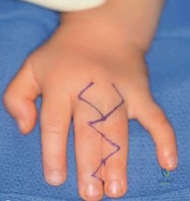
Structure the answer into 1) Exposure via curvilinear incision along the hypothenar border. 2) Ulnar nerve decompression and neurolysis. 3) Resection of the pathological artery segment. 4) Reconstruction with a reversed autogenous vein graft (e.g., cephalic vein) to restore flow to the palmar arch. 5) Use of microvascular techniques and post-operative antiplatelet therapy.
