Lumbrical Plus Deformity: Epidemiology, Anatomy, and Pathophysiology
Key Takeaway
Lumbrical plus deformity is an uncommon hand condition where attempted finger flexion paradoxically extends the interphalangeal (IP) joints. It often arises as an iatrogenic complication following flexor tendon surgery, due to over-tensioning of FDP repairs or transfers, or FDP avulsion. This disrupts normal force transmission, causing the lumbrical muscle to act as an IP joint extensor.
A 35-year-old male presents with a chief complaint of "difficulty making a fist" following a primary zone II flexor tendon repair 4 months ago. On examination, when you ask the patient to actively flex his fingers, the MCP joint of the ring finger flexes normally, but the PIP and DIP joints extend paradoxically. What is the clinical diagnosis, and what is the underlying biomechanical pathophysiology?
Candidate: The patient has a lumbrical plus deformity. This happens because the FDP tendon repair is too tight or the tendon has retracted. When the patient tries to flex the finger, the FDP muscle pulls on the lumbrical instead of the distal phalanx. This causes the lumbrical to extend the PIP and DIP joints while the MCP is being flexed by the lumbrical’s other action.
Failing to mention the specific mechanical disadvantage: the FDP excursion is limited, forcing the force of muscle contraction to be transmitted through the lumbrical origin rather than the distal phalanx. Also, candidates often forget to differentiate this from a stiff PIP joint or "quadriga effect" in the exam.
This is a Lumbrical Plus Deformity. Pathophysiologically, it results from a relative mismatch between FDP tension and excursion. When the FDP is tethered or over-tightened, the FDP muscle contraction cannot achieve the excursion required for DIP flexion. Instead, the force is transmitted proximally to the origin of the lumbrical muscle. The lumbrical, acting as a bipennate muscle originating from the FDP, then exerts its primary action—MCP flexion and IP extension—paradoxically, resulting in the observed IP extension during attempted active flexion.
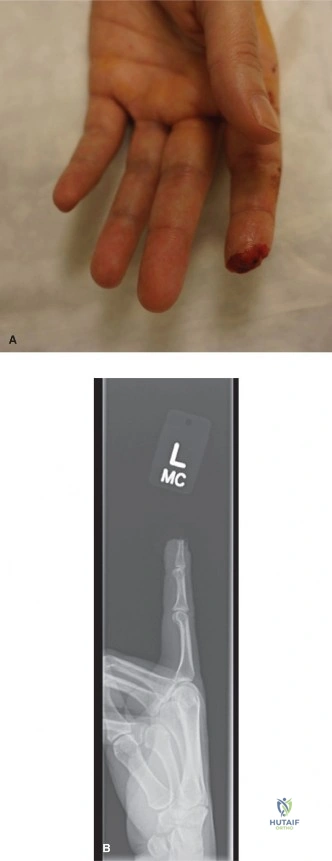
The patient is booked for surgery. Describe your intraoperative decision-making process and the procedure you would perform to correct this. Use the provided image to explain the surgical logic if relevant.
Candidate: I would perform a lumbrical tenotomy. I would identify the lumbrical origin on the radial side of the FDP and transect it. If the FDP is still too tight, I would perform a Z-lengthening of the FDP in the palm or forearm as shown in the diagram.
Starting with FDP lengthening. The Gold Standard is to start with the simplest, least morbid procedure (tenotomy). Furthermore, failing to mention intraoperative "tension testing" to confirm if the tenotomy alone resolves the paradoxical extension is a major oversight.
The first-line surgical treatment is a lumbrical tenotomy. I would approach via a volar zigzag (Brunner) incision, protect the neurovascular bundles, and release the lumbrical tendon from its origin on the FDP. Crucially, I would perform an intraoperative assessment; if paradoxical extension persists, it confirms that the FDP itself is significantly over-tensioned. In that specific scenario, I would proceed to an FDP Z-lengthening (as illustrated in the provided image). The goal is to restore the digital cascade and ensure smooth FDP excursion without compromising the A2/A4 pulleys.
