Diagnostic Approach to Median Nerve Schwannoma: A Detailed Orthopedic Case Study
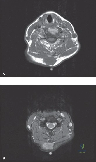
Key Takeaway
Median nerve schwannoma diagnosis involves progressive pain, paresthesia, and a palpable forearm mass, often with a positive Tinel's sign. Clinical examination reveals neurological deficits. MRI is key, demonstrating a characteristic 'target sign' on T2-weighted images and avid, heterogeneous post-contrast enhancement, confirming the nerve's involvement and guiding further management.
A 48-year-old man presents with a 10-month history of progressive volar forearm pain and paresthesia in the median nerve distribution. On examination, you identify a 3cm firm mass in the distal forearm that is mobile transversely but restricted longitudinally. What is the most likely diagnosis, and what is the significance of this physical examination finding?
Candidate: The most likely diagnosis is a schwannoma of the median nerve. The mobility pattern—mobile transversely but fixed longitudinally—is the hallmark clinical sign of an intraneural tumor, as it is tethered to the nerve of origin.
Failing to mention the specific mobility pattern or simply calling it a "soft tissue mass." Candidates often miss the link between the clinical sign and the underlying anatomy (nerve origin), which is essential to distinguish it from extraneural masses like a lipoma or ganglion.
The patient likely has a benign peripheral nerve sheath tumor, specifically a schwannoma. The "transverse mobility vs. longitudinal restriction" sign confirms the lesion is intraneural. I would also note the positive Tinel's sign. I would differentiate this from a neurofibroma by noting that schwannomas are typically eccentric and encapsulated, whereas neurofibromas are central and infiltrate the fascicles.
You obtain an MRI of the forearm. Describe the expected findings for this lesion and explain the significance of the "split-fat sign."
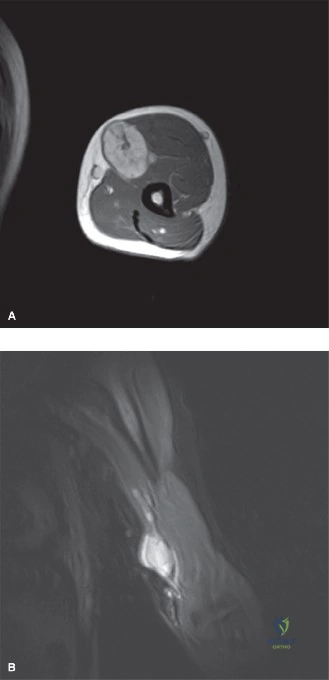
Candidate: On MRI, I expect a T2 hyperintense mass. The "target sign" is common, showing a peripheral rim of high signal and a central low signal core. The "split-fat sign" is a rim of adipose tissue at the poles of the mass, which indicates the lesion is contained within the nerve sheath (intraneural).
Ignoring the "target sign" or failing to explain the pathology behind it (Antoni A vs Antoni B tissue). Simply listing the signs without explaining that the split-fat sign helps differentiate intraneural from extraneural masses shows a lack of deep diagnostic understanding.
I would expect T1 isointensity and T2 hyperintensity. The 'target sign' (peripheral myxoid Antoni B, central cellular Antoni A) is highly characteristic. The 'split-fat sign'—a crescent of fat at the tumor's proximal and distal ends—is pathognomonic for an intraneural origin. I would also look for the 'fascicular sign,' which shows the splayed nerve fascicles on the tumor periphery.
The patient requests a core needle biopsy prior to surgery to confirm the diagnosis. How do you respond?
Candidate: I would strongly advise against a core needle biopsy. It carries a significant risk of iatrogenic fascicular injury and can induce severe neuropathic pain. The diagnosis is strongly supported by the clinical presentation and MRI findings; therefore, the biopsy is unnecessary and potentially harmful.
Agreeing to a biopsy or being unsure. Examiners need you to be decisive: for suspected benign nerve sheath tumors, biopsy is contraindicated due to the high risk of permanent nerve damage.
I would explain that a core needle biopsy is contraindicated in this scenario. The risk of permanent neurological deficit outweighs the diagnostic value. In orthopaedic oncology, we prioritize the 'imaging-clinical correlation.' If the imaging is pathognomonic for a benign schwannoma, we proceed to surgical enucleation as both the diagnostic and definitive therapeutic step.
You are in the operating theatre. You have identified the median nerve and see the mass. Describe your surgical technique for enucleation.
Candidate: I would use microsurgical techniques, preferably with an operating microscope. After identifying the nerve in healthy tissue proximally and distally, I would perform a longitudinal epineurotomy in an avascular zone and carefully dissect the functional fascicles away from the tumor capsule to enucleate the mass.
Forgetting to identify the nerve in "virgin" (unaffected) tissue first, or failing to mention the use of magnification. Simply saying "I'll take it out" ignores the crucial principle of protecting functional fascicles.
I follow a structured microsurgical approach: 1. Identify the nerve in proximal and distal healthy tissue. 2. Use longitudinal epineurotomy in an avascular zone. 3. Use blunt dissection to isolate the tumor capsule from splayed fascicles. 4. Use intraoperative nerve stimulation to ensure the fascicle of origin is non-conductive/non-functional before sacrificing it. 5. Avoid closing the epineurium to prevent post-op nerve compression from hematoma.
