Erosive Osteoarthritis of the Index Finger PIP: Comprehensive Anatomy, Biomechanics & Epidemiology
Key Takeaway
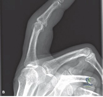
Erosive osteoarthritis (EOA) of the index finger PIP is an aggressive inflammatory form of OA, predominantly affecting middle-aged women. It causes rapid cartilage loss and subchondral bone erosions, often leading to a 'gull-wing' deformity. This significantly disrupts the joint's complex anatomy and biomechanics, resulting in pain, stiffness, and functional impairment.
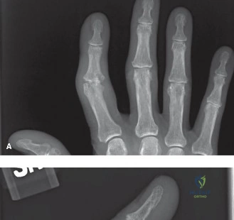
A 55-year-old female presents with a 2-year history of painful, swollen, and stiff proximal interphalangeal (PIP) joints of her fingers. She reports intermittent "flares" of redness and intense pain. Clinical examination reveals characteristic "gull-wing" deformity on radiographs. Describe the pathophysiology of this condition and the key clinical features that distinguish it from primary osteoarthritis.
Candidate: This is Erosive Osteoarthritis (EOA). Unlike primary OA, which is purely degenerative, EOA involves an inflammatory phase with synovial proliferation and cytokine-mediated cartilage degradation. Clinically, it presents with episodic inflammation, more rapid joint destruction, and classic "gull-wing" central erosions on X-ray. It typically affects post-menopausal women.
Failing to mention the episodic inflammatory nature or confusing the radiographic findings with Rheumatoid Arthritis. A poor candidate might just say "it's bad arthritis" without highlighting the specific central erosive pattern that defines EOA.
Structure the answer by defining EOA as an inflammatory phenotype of hand OA. Mention the "Gull-wing" deformity (central erosions with peripheral osteophytes), the patient demographic (female-to-male ratio 5:1 to 12:1), and the specific pathophysiology: synovial hypertrophy causing active chondrolysis. Explicitly state the differential diagnosis: it must be distinguished from RA or Psoriatic arthritis via serum inflammatory markers (typically negative in EOA).
The patient has failed 6 months of conservative management for her index PIP joint. She is a seamstress and needs her finger for fine motor tasks. You are considering surgery. How would you counsel her on the choice between arthrodesis and arthroplasty, and what is your preferred technique for the index finger?
Candidate: Arthrodesis provides stability and pain relief, which is ideal for the index finger due to high pinch demands. Arthroplasty offers motion but carries risks of implant failure or instability. For a seamstress, I would likely recommend arthrodesis in 30-45 degrees of flexion, as it ensures the stability required for her occupation.
Ignoring the occupational functional demands. A candidate who suggests arthroplasty for a high-demand, high-pinch finger without discussing the stability-vs-motion trade-off will likely score lower.
Categorize the decision based on Functional Demands vs. Joint Biology. State clearly: Arthrodesis is the gold standard for the index PIP in patients requiring forceful pinch/stability. For Arthroplasty (e.g., Pyrocarbon), specify it is reserved for patients prioritizing motion over strength, with good bone stock. Mention the optimal fusion angle (30-45 degrees) and emphasize that for a seamstress, the predictability of arthrodesis is superior to the variable outcomes of arthroplasty in the index finger.
You have decided to perform an arthrodesis. Looking at this intraoperative image, describe the fixation strategy and why you have chosen this configuration.
Candidate: The image shows a dorsal mini-fragment plate used for PIP fusion. This provides rigid internal fixation, allowing for early mobilization of the adjacent MCP and DIP joints, which prevents overall hand stiffness and promotes better union rates compared to simple K-wire fixation.
Suggesting that the goal of the surgery is to keep the entire finger immobilized. Failing to emphasize the importance of early motion in the *adjacent* joints (MCP/DIP) is a significant oversight.
Frame the answer around Rigid Internal Fixation (RIF). Explain that a dorsal mini-plate (1.5/2.0mm) is superior to K-wires for achieving compression across the arthrodesis site. Mention the "tension-band" effect of the plate, the reduction in non-union rates, and the critical post-operative protocol: immobilization of the PIP joint while strictly encouraging active/passive motion of the MCP and DIP joints to prevent digit-wide morbidity.
