Comprehensive Case Study: Chronic Exertional Compartment Syndrome in a Long-Distance Runner
Key Takeaway
Chronic Exertional Compartment Syndrome (CECS) in runners is diagnosed through a combination of classic exertional leg pain, specific clinical findings post-exertion (weakness, paresthesia), and ruling out other pathologies with imaging. The gold standard for definitive diagnosis involves invasive compartment pressure testing performed during or immediately after symptomatic exercise.
A 25-year-old elite long-distance runner presents with a 6-month history of bilateral lower leg "bursting" pain and paresthesia. Symptoms occur consistently 10 minutes into runs and resolve at rest. You suspect Chronic Exertional Compartment Syndrome (CECS). What is your structured approach to the diagnosis and management of this patient?
Candidate: I would take a thorough history to confirm the exertional nature and rule out stress fractures or arterial issues. I'd perform a physical exam, which is often normal at rest. To diagnose, I would order dynamic intracompartmental pressure monitoring using the Pedowitz criteria. If confirmed and conservative management fails, I would discuss a fasciotomy.
Candidates often forget the "Red Flag" differentials. Failing to explicitly mention ruling out Popliteal Artery Entrapment Syndrome (PAES) or stress fractures makes the answer incomplete. Furthermore, simply saying "fasciotomy" without mentioning the surgical technique (e.g., fasciectomy to reduce recurrence) shows a lack of senior-level clinical reasoning.
A structured response is essential: 1. Clinical Presentation: Emphasize the "bursting" nature and the predictable symptom-free interval. 2. Differential Diagnosis: Systematically exclude Medial Tibial Stress Syndrome (MTSS), Tibial Stress Fractures, PAES, and nerve entrapment (superficial peroneal). 3. Objective Testing: State the Pedowitz criteria (Resting ≥15, 1-min post ≥30, or 5-min post ≥20 mmHg). 4. Management: Address the failure of conservative treatment (>80% failure in elite athletes), the role of partial fasciectomy vs. simple fasciotomy to mitigate recurrence, and the critical importance of protecting the superficial peroneal nerve during surgery.
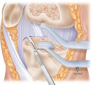
Consider the image below, which represents the surgical field during an anterolateral release. What are the critical structures at risk, and how do you ensure a safe, definitive release?
Candidate: The primary structure at risk is the superficial peroneal nerve. I would identify it as it pierces the deep fascia of the lateral compartment. I would perform an open, longitudinal incision, perform a partial fasciectomy, and ensure complete proximal to distal release, checking for hemostasis after tourniquet deflation.
Missing the anatomical variation of the superficial peroneal nerve. Candidates who assume the nerve is always in the same spot risk iatrogenic injury. Also, failing to mention the necessity of decompressing both the anterior and lateral compartments if pressure testing indicates multi-compartment involvement is a frequent oversight.
Start with the anatomy: the superficial peroneal nerve pierces the lateral fascia ~10-12cm proximal to the lateral malleolus but is highly variable. - Exposure: Longitudinal incision over the intermuscular septum allows access to both compartments. - Technique: Use a partial fasciectomy (1-2cm strip) to prevent fascial re-approximation—a common cause of recurrence. - Safety: Mobilize the nerve, release proximal to the tibial tubercle, and distal to the extensor retinaculum. - Hemostasis: Mandatory tourniquet release and bipolar cautery to prevent postoperative hematoma and subsequent fibrosis.
