Acute Compartment Syndrome Diagnosis in Open Tibial Fractures: A Clinical Case Study

Key Takeaway
Acute compartment syndrome after tibial fracture presents with pain disproportionate to injury, unremitting despite analgesia. Key red flags include tense, 'wood-hard' compartments, severe pain on passive stretch, progressive neurological deficits (weakness, paresthesia), and evolving vascular compromise (diminished pulses, delayed capillary refill). Early recognition is vital.
You are the registrar on call in the trauma unit. A 28-year-old male arrives following a 70 km/h motor vehicle collision with a mid-distal tibial fracture. He is complaining of intractable, worsening leg pain despite 10mg morphine and 50mcg fentanyl. On examination, the leg is tense, and he has pain on passive stretch of the toes. Distal pulses are present. What is your immediate clinical concern, and how do you differentiate this from "routine" fracture pain?
Candidate: My immediate concern is acute compartment syndrome (ACS). I would differentiate it from normal fracture pain by the fact that the pain is "out of proportion" to the injury, is not relieved by analgesia or splinting, and is exacerbated by passive stretch of the involved muscle groups. The tense, "wood-hard" compartments are a key clinical finding, and I would be careful not to rely on the presence of pulses, as they are a very late sign.
Candidates often focus too heavily on the "5 Ps" (specifically pulselessness). A failing answer suggests that because pulses are present, ACS is excluded. Furthermore, failing to mention the specific test of "pain on passive stretch" as the most sensitive clinical indicator is a major oversight.
The candidate must explicitly state that ACS is a clinical diagnosis. Highlight that pain out of proportion to injury and pain on passive stretch are the most reliable clinical markers. Emphasize that peripheral pulses remain intact until intra-compartmental pressure exceeds systolic blood pressure, making it a late and unreliable sign. A top-tier response mentions the "Delta P" (Diastolic BP - Compartment Pressure) as the objective threshold (<30 mmHg) for surgical intervention if the clinical picture is equivocal.
You review the radiographs of this patient, showing an OTA/AO 42-B3 fracture. You note a 2cm open wound. Does the presence of this open wound exclude the possibility of compartment syndrome? How does this influence your surgical planning?
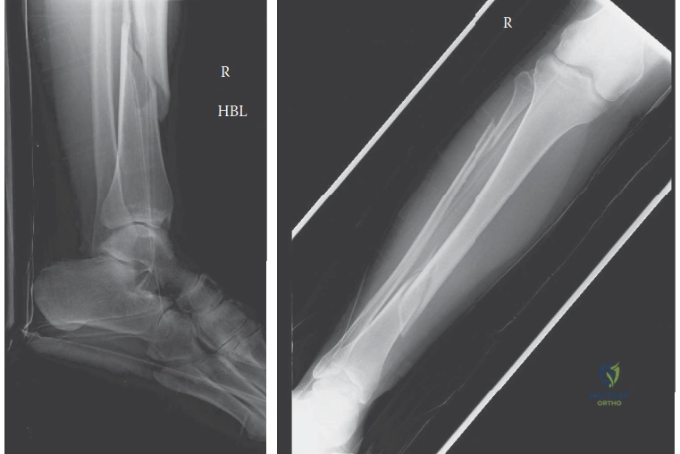
Candidate: The open wound does not exclude compartment syndrome. In fact, studies show open fractures can have a similar or higher incidence of ACS because the fascial defect is rarely large enough to provide adequate decompression. I would proceed with an emergent four-compartment fasciotomy. For the fracture itself, I would favor an unreamed intramedullary nail to provide stability, avoiding reaming to prevent further pressure elevation, and manage the wounds with vacuum-assisted closure.
Assuming the fracture is "open enough" to self-decompress is a catastrophic error. Candidates often fail to address the risk of using reamed nails in this context, which can increase intramedullary pressure and potentially exacerbate the compartment syndrome.
State clearly that open fractures are NOT decompressed by the injury. Outline the surgical strategy: Four-compartment fasciotomy via dual incisions (Mubarak and Owen) for safety and thoroughness. Confirm that reaming is contraindicated (use unreamed IM nail) to prevent further pressure increase, and prioritize negative pressure wound therapy (NPWT) for the fasciotomy sites, ensuring they are left open as primary closure is contraindicated.
During your four-compartment fasciotomy, which anatomical structures are at the highest risk of iatrogenic injury during the release of the lateral and deep posterior compartments?
Candidate: When releasing the lateral compartment, the common peroneal nerve as it winds around the fibular neck is the critical structure at risk. When releasing the deep posterior compartment, the posterior tibial neurovascular bundle (posterior tibial artery, vein, and tibial nerve) must be identified and protected, particularly as the deep fascia is released.
Forgetting the superficial peroneal nerve, which exits the lateral fascia in the distal third of the leg, or failing to mention the avoidance of the saphenous nerve and vein during the posteromedial incision.
Systematically list the structures: 1. **Lateral compartment:** Common peroneal nerve (proximally at fibular neck) and superficial peroneal nerve (distally as it pierces fascia). 2. **Posterior compartments:** The neurovascular bundle (Posterior Tibial A/V/N) in the deep posterior compartment. 3. **Posteromedial approach:** The saphenous vein and nerve, which are at risk if the incision is placed too anteriorly.
