Pediatric Trigger Thumb (Stenosing Tenosynovitis): A Comprehensive Academic Guide
Key Takeaway
Pediatric trigger thumb is FPL stenosing tenosynovitis, where a Notta's nodule catches at the A1 pulley, causing locking or triggering. It's developmental, not congenital. Treatment ranges from observation in young children to surgical A1 pulley release for persistent locking or a fixed flexion deformity, preventing secondary joint changes.
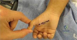
A mother presents with her 18-month-old child, concerned about a "clicking" sensation in the left thumb. On examination, you notice the IP joint is locked in 30 degrees of flexion, and there is a palpable nodule at the volar MCP crease. What is your diagnosis, and how do you justify the timing of your management decision?
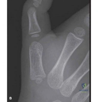
Candidate: The diagnosis is a pediatric trigger thumb, caused by the entrapment of the flexor pollicis longus tendon at the A1 pulley, often associated with a Notta’s nodule. At 18 months, I would discuss the natural history. Many of these resolve, but since the thumb is now fixed in flexion, I would consider a trial of extension splinting or discuss surgical release if the parents prefer or if it remains locked.
The candidate fails to acknowledge the "acquired" nature of the condition, wrongly calling it "congenital." They also often suggest immediate surgery without referencing the evidence (e.g., Dinham and Meggitt) that a significant proportion of children, especially those younger than 2 years, may still show spontaneous resolution or benefit from non-operative measures before committing to a general anesthetic.
State the diagnosis as an "acquired stenosing tenosynovitis of the FPL." Support the management by citing the natural history: observation is valid for children <12-24 months if the deformity is flexible. However, because this patient has a "fixed flexion contracture," the prognosis for spontaneous resolution is reduced. Mention a structured approach: initial trial of splinting/stretching, and if unsuccessful after 3-6 months—or if the child remains locked—surgical A1 pulley release is indicated.
You have decided to proceed with an A1 pulley release for this child. You are now in the operating theatre. Describe your surgical approach, specifically focusing on the neurovascular anatomy and how you avoid iatrogenic injury.
Candidate: I would use a transverse incision at the MCP flexion crease. After skin incision, I perform blunt dissection to find the A1 pulley. I must watch for the radial digital nerve, which crosses obliquely. I will use magnification, release the A1 pulley longitudinally, but ensure I do not cut the oblique pulley, which is essential for preventing bowstringing.
Candidates often forget to mention the use of a tourniquet for a bloodless field, which is critical for identifying these tiny nerves in a child. They also frequently fail to explicitly mention identifying the *oblique* pulley to preserve it, which is the most common technical error leading to long-term morbidity (bowstringing).
Structure the response: 1. **Setup:** Tourniquet for a bloodless field and loupe magnification. 2. **Incision:** Transverse at the MCP crease. 3. **Dissection:** Strictly blunt longitudinal spreading to reach the A1 pulley. 4. **Anatomical landmark:** Explicitly identify the radial digital nerve, noting its oblique course from ulnar to radial. 5. **Technique:** Longitudinal release of A1; preserve the oblique pulley to prevent FPL bowstringing. 6. **Verification:** Test passive IP joint motion intra-operatively to confirm smooth glide.
Post-operatively, you notice the child has persistent IP joint stiffness despite a perfect release of the A1 pulley. How do you manage this, and what are the potential long-term complications if the release is not managed correctly?
Candidate: Stiffness is likely due to adaptive shortening of the volar structures from chronic disuse. I would start active and passive motion exercises, potentially with night splinting. Complications to look out for include recurrence from an incomplete release or, worse, iatrogenic nerve injury or tendon bowstringing if the oblique pulley was violated.
The candidate ignores the "rehabilitation" aspect, failing to suggest parental involvement in stretching. They also often miss the distinction between *incomplete release* (requiring revision) and *joint contracture* (requiring physical therapy), treating them as the same problem.
Differentiate between mechanical blockage and physiological contracture. If the release was verified intra-operatively, the stiffness is likely an adaptive contracture of the volar plate/collateral ligaments. Management: Guided parent-led passive stretching and early active motion. If unsuccessful, add night extension splinting. If the stiffness were mechanical (clicking persists), this represents an incomplete release requiring revision. Highlight the "Red Flag" of bowstringing (loss of flexion power) as a permanent, avoidable complication requiring reconstructive surgery.
