Complex Index Finger MCP & PIP Joint Injuries: Diagnosis, Surgical Anatomy, and Management

Key Takeaway
Complex index finger MCP and PIP joint injuries involve intricate damage to articular surfaces, ligaments, and soft tissues from high-energy trauma. Accurate diagnosis, vital for preserving hand function, necessitates understanding their epidemiology, detailed surgical anatomy, and biomechanics. Meticulous management prevents chronic pain and functional impairment.
A 28-year-old rugby player presents with a painful, swollen index finger following an axial load injury. You are presented with the following radiograph. How do you classify this, and what is your immediate assessment of the stability?
Candidate: The radiograph shows an intra-articular fracture-dislocation of the PIP joint. I would look at the articular surface involvement, check for subluxation on the lateral view, and perform a clinical stress test to assess for collateral ligament integrity.
Focusing only on the fracture line without mentioning the "pilon" nature of the injury or failing to address the integrity of the volar plate and collateral ligaments. Candidates often forget to mention the need for a CT scan to assess the percentage of articular surface involvement.
This is a dorsal fracture-dislocation of the PIP joint. My assessment involves: 1) Determining the percentage of the articular surface involved (typically >30% implies instability). 2) Evaluating for dorsal subluxation of the middle phalanx. 3) Checking for avulsion of the volar plate. I would request a CT scan to define the comminution. Clinically, I must ensure there is no concomitant central slip injury, as this would indicate a high risk of Boutonnière deformity.
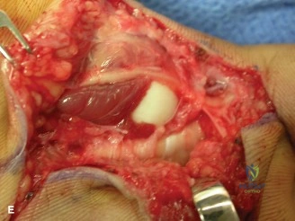
During the procedure, you encounter a fracture of the proximal phalanx condyle. The fragment is small but clearly intra-articular. How do you approach the decision-making for fixation?
Candidate: I would aim for anatomical reduction. If the fragment is large enough, I would use a 1.0 or 1.3mm headless compression screw. If it is too small, I might use K-wires or a tension band technique.
Failing to mention the protection of the neurovascular bundles or the risk of rotational malalignment, which is the most common cause of a poor functional outcome in condylar fractures.
The priority is anatomical restoration to prevent post-traumatic arthritis and restoring rotational alignment to prevent "scissoring." I would use a mid-axial approach to protect the collateral ligaments. Fixation choice is guided by fragment size: headless compression screws (1.0/1.3mm) provide the best compression, but if too comminuted, a tension band wire or K-wires may be safer. I would confirm the reduction fluoroscopically in multiple planes, specifically looking for rotational alignment against the adjacent digits.
What are the relative indications for surgical versus non-operative management in complex PIP joint injuries, and how does the concept of "position of safety" apply post-operatively?
Candidate: Surgery is for unstable or irreducible injuries. Non-operative is for stable, congruent joints. The position of safety is MCP flexion and PIP/DIP extension.
The candidate fails to explain *why* the position of safety is used (collateral ligament length) and misses the nuance that PIP joint protocols often differ from MCP joint protocols regarding flexion/extension.
Surgery is indicated for intra-articular step-off >1-2mm, irreducible dislocations, or instability on stress testing. The "position of safety" (MCP flexion ~70-90°, IP joints neutral) is used to keep the collateral ligaments taut, preventing contracture. However, in PIP joint fracture-dislocations, the position of immobilization must be carefully balanced—often using an extension block splint to maintain reduction while allowing gradual flexion, as the risk of PIP joint stiffness is extremely high compared to the MCP joint.
