Broken Radius Fractures: What You Need to Know About Your Broken Wrist
Key Takeaway
Learn more about Broken Radius Fractures: What You Need to Know About Your Broken Wrist and how to manage it. Distal radius fractures occur when the radius, one of the two forearm bones located on the thumb side, breaks close to the wrist. These **radius fractures broken** about an inch from the bone's end are very common, often resulting from falls onto an outstretched arm. Fractures can be intra-articular, extra-articular, open, or comminuted, with Colles fracture being a common type.
Introduction and Epidemiology
Distal radius fractures represent one of the most frequently encountered osseous injuries in orthopedic trauma, accounting for approximately one sixth of all fractures treated in emergency departments. The epidemiologic profile of these fractures demonstrates a classic bimodal distribution. In younger demographics, these injuries typically result from high energy trauma, such as motor vehicle collisions, falls from significant heights, or athletic injuries, often presenting with complex, intra articular comminution and associated soft tissue or neurovascular compromise. Conversely, in the elderly population, distal radius fractures predominantly occur secondary to low energy mechanisms, most notably a fall from a standing height onto an outstretched hand. This latter cohort is heavily influenced by decreased bone mineral density, making the distal radius a sentinel fracture for osteoporosis.
The historical eponyms associated with distal radius fractures—Colles, Smith, Barton, and Chauffeur—while clinically ubiquitous, have largely been superseded in academic discourse by standardized, morphologic classification systems that guide surgical decision making. The Orthopaedic Trauma Association and Arbeitsgemeinschaft für Osteosynthesefragen classification system remains the most comprehensive, categorizing fractures into extra articular, partial articular, and complete articular patterns. Furthermore, the Fernandez classification provides a mechanistic framework, categorizing fractures based on bending, shearing, compression, avulsion, or combined forces, which directly informs the requisite biomechanical strategy for surgical fixation.
Understanding the complex interplay between fracture morphology, patient physiology, and functional demands is paramount. The evolution of fracture management has shifted significantly over the past two decades, moving from predominantly closed reduction and cast immobilization or external fixation toward open reduction and internal fixation utilizing volar locking plate technology. This paradigm shift necessitates a rigorous understanding of distal radius anatomy, biomechanics, and surgical techniques to optimize patient outcomes and minimize iatrogenic complications.
Surgical Anatomy and Biomechanics
Osteology and Radiographic Parameters
The distal radius is a complex three dimensional structure that transitions from the diaphyseal triangular cross section to a broad, flat metaphyseal flare. This geometric expansion is characterized by a relatively thin cortical shell surrounding a region of dense cancellous bone, which is highly susceptible to compressive failure during axial loading.
Restoration of native radiographic parameters is critical for functional recovery. The normal distal radius exhibits a radial inclination averaging 22 to 24 degrees, a palmar tilt of 11 to 12 degrees, and a radial height of 11 to 12 millimeters. Ulnar variance, the relative length of the distal radius to the distal ulna, should ideally be neutral, though it varies slightly among individuals. Alterations in these parameters significantly disrupt radiocarpal and distal radioulnar joint kinematics. For instance, dorsal angulation exceeding 20 degrees shifts the load bearing axis dorsally, increasing the load on the distal ulna and the triangular fibrocartilage complex, leading to ulnocarpal impaction syndrome and restricted forearm rotation.
Articular Anatomy
The articular surface of the distal radius is divided into three distinct fossae. The scaphoid fossa is triangular and located radially, separated from the lunate fossa by the interfossal ridge. The lunate fossa is quadrilateral and situated ulnarly. The sigmoid notch, located on the ulnar aspect of the distal radius, articulates with the ulnar head to form the distal radioulnar joint. The congruity of the sigmoid notch is vital for pronation and supination; intra articular step offs or gaps exceeding 2 millimeters in this region or the radiocarpal articular surface dramatically increase the risk of post traumatic osteoarthritis.
Soft Tissue and Ligamentous Constraints
The stability of the radiocarpal and midcarpal joints relies heavily on extrinsic and intrinsic ligamentous complexes. The volar radiocarpal ligaments, including the radioscaphocapitate, long radiolunate, and short radiolunate ligaments, are stout structures that provide primary volar stability. The dorsal radiocarpal ligament provides secondary restraint. The triangular fibrocartilage complex is the primary stabilizer of the distal radioulnar joint and serves as a load bearing structure for the ulnar carpus.
The Watershed Line
From a surgical perspective, the volar aspect of the distal radius features a critical landmark known as the watershed line. This is a transverse bony ridge situated near the articular margin that separates the flat pronator quadratus fossa from the distal articular surface. The flexor pollicis longus and flexor digitorum profundus tendons glide directly over this ridge. Volar plates placed distal to the watershed line risk prominent hardware impinging on these flexor tendons, leading to tenosynovitis or catastrophic attrition rupture.
Indications and Contraindications
The decision to proceed with operative intervention versus non operative management hinges on fracture stability, articular congruity, and the functional demands of the patient. Lafontaine previously established criteria for instability, which include initial dorsal tilt greater than 20 degrees, dorsal comminution, intra articular extension, associated ulnar fracture, and advanced patient age. Fractures exhibiting these characteristics are highly likely to displace secondarily despite adequate initial closed reduction and cast immobilization.
| Parameter | Non Operative Management | Operative Management |
|---|---|---|
| Radial Shortening | Less than 3 mm | Greater than 3 mm |
| Dorsal Tilt | Less than 10 degrees past neutral | Greater than 10 degrees past neutral |
| Intra Articular Step Off | Less than 2 mm | Greater than or equal to 2 mm |
| Volar Tilt | Acceptable if minimal displacement | Volar displacement or Smith variant |
| Fracture Stability | Stable post closed reduction | Loss of reduction in cast |
| Soft Tissue Status | Closed, intact neurovascular status | Open fracture, acute carpal tunnel syndrome |
| Concomitant Injuries | Isolated injury | Polytrauma, floating elbow, DRUJ instability |
| Patient Factors | Low demand, significant comorbidities | High functional demand, physiologically young |
Absolute and Relative Contraindications
Absolute contraindications to surgical intervention include active local or systemic infection, severe medical comorbidities precluding the administration of anesthesia, and non ambulatory patients with minimal upper extremity functional demands. Relative contraindications include severe osteopenia where hardware purchase may be compromised, although modern locking plate technology has largely mitigated this concern. Severe baseline dementia or an inability to comply with post operative rehabilitation protocols also represent relative contraindications, as non compliance can lead to hardware failure or severe stiffness.
Pre Operative Planning and Patient Positioning
Advanced Imaging and Templating
Standard posteroanterior, lateral, and oblique radiographs of the wrist are mandatory. The posteroanterior view assesses radial height, radial inclination, and ulnar variance. The lateral view is critical for evaluating palmar tilt and carpal alignment. The teardrop angle, representing the volar rim of the lunate fossa, should be scrutinized on the lateral radiograph to assess for volar rim fractures.
For complex intra articular fractures, a non contrast computed tomography scan is highly recommended. Computed tomography allows for the precise characterization of articular comminution, identification of central die punch fragments, and evaluation of the sigmoid notch and volar rim. Three dimensional reconstructions can aid in spatial conceptualization and preoperative templating. Templating involves selecting the appropriate plate size, determining screw trajectories to ensure subchondral support without intra articular penetration, and anticipating the need for supplemental fixation such as fragment specific plates or Kirschner wires.
Operating Room Setup and Patient Positioning
The patient is positioned supine on the operating table with the operative extremity extended onto a radiolucent hand table. A non sterile tourniquet is applied to the proximal arm. Regional anesthesia, typically an axillary or supraclavicular brachial plexus block, is preferred as it provides excellent intraoperative muscle relaxation and superior post operative analgesia, though general anesthesia may be utilized based on patient preference or contraindications to regional blockade.
Fluoroscopy is essential. The C arm is typically brought in from the head of the table or parallel to the hand table, perpendicular to the long axis of the patient. The monitor should be positioned directly across from the surgeon for ergonomic visualization. Prior to prepping and draping, the surgeon must confirm that adequate posteroanterior, lateral, and oblique fluoroscopic images can be obtained without obstruction.
Detailed Surgical Approach and Technique
The Volar Henry Approach
The modified volar Henry approach is the workhorse exposure for volar plating of distal radius fractures. It utilizes the internervous plane between the median nerve innervated flexor carpi radialis and the radial nerve innervated brachioradialis.
Superficial Dissection
A longitudinal incision is made over the course of the flexor carpi radialis tendon, extending approximately 8 to 10 centimeters proximally from the wrist crease. The subcutaneous tissue is sharply divided. Care must be taken to identify and protect the palmar cutaneous branch of the median nerve, which typically lies ulnar to the flexor carpi radialis tendon but can have variable arborization. The flexor carpi radialis tendon sheath is incised longitudinally. The tendon is retracted ulnarly, which inherently protects the median nerve and the palmar cutaneous branch.
Deep Dissection and Exposure
The floor of the flexor carpi radialis sheath is incised to reveal the flexor pollicis longus muscle belly and tendon. The flexor pollicis longus is retracted ulnarly to expose the pronator quadratus muscle. At the radial aspect of the wound, the radial artery is identified and gently retracted radially.
The brachioradialis tendon insertion on the radial styloid is a major deforming force. Releasing the distal aspect of the brachioradialis from its insertion is a critical step; it facilitates mobilization of the radial styloid fragment and improves visualization of the distal radius. The pronator quadratus is then elevated off the radius via an L shaped incision along its radial and distal borders. A small cuff of muscle may be left distally and radially for later repair, though evidence suggests that formal repair of the pronator quadratus does not significantly alter functional outcomes. Elevation of the pronator quadratus exposes the volar cortex of the distal radius and the fracture site.
Fracture Reduction Techniques
Hematoma and interposed soft tissue are debrided from the fracture site. Reduction is achieved through a combination of longitudinal traction, ligamentotaxis, and direct manipulation. For dorsally displaced fractures, the surgeon volar flexes the distal fragment while applying traction.
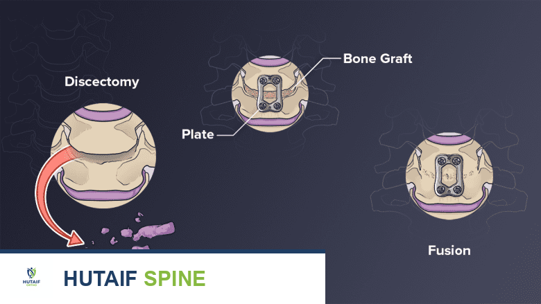
Intrafocal pinning, utilizing the Kapandji technique, can be employed temporarily to lever the fracture into anatomic alignment. Alternatively, Kirschner wires can be inserted into the distal fragments to act as joysticks for manipulation. Central die punch fragments may require elevation through the fracture window and bone grafting (autograft, allograft, or synthetic bone substitutes) to support the articular surface.
Plate Application and Fixation
Once provisional reduction is achieved and confirmed fluoroscopically, the volar locking plate is applied. The plate must be positioned proximal to the watershed line to prevent flexor tendon irritation. A cortical screw is typically placed in the elongated gliding hole of the proximal shaft to secure the plate to the diaphysis, allowing for proximal distal adjustments.
Distal fixation relies on locking screws that act as a fixed angle construct, providing subchondral support to the articular surface. The trajectory of these screws is critical; they must be directed dorsally and slightly proximally to avoid intra articular penetration. Fluoroscopic evaluation using specialized views, such as the dorsal tangential view, is mandatory to confirm that screws have not breached the dorsal cortex, which could lead to extensor tendon rupture.
Alternative Approaches and Fragment Specific Fixation
While volar plating addresses the majority of distal radius fractures, specific patterns require alternative strategies. Severe dorsal comminution or dorsal Barton fractures may necessitate a dorsal approach. This typically involves an incision over the third dorsal compartment, transposition of the extensor pollicis longus tendon, and elevation of the second and fourth compartment contents to expose the dorsal radius.
For complex, multi fragmentary articular fractures, fragment specific fixation may be required. This involves the use of low profile, specialized plates (such as radial pin plates, dorsal delta plates, or volar hook plates) to capture individual articular fragments that cannot be adequately secured by a single standard volar plate. In cases of severe soft tissue compromise or extreme comminution precluding internal fixation, a spanning external fixator combined with percutaneous pinning remains a viable salvage option.
Complications and Management
Despite advancements in surgical technique and implant design, complications following distal radius fracture fixation remain a significant clinical challenge. Thorough preoperative planning and meticulous surgical execution are required to mitigate these risks.
| Complication | Estimated Incidence | Etiology | Salvage and Management Strategy |
|---|---|---|---|
| Extensor Tendon Rupture | 1 to 5 Percent | Prominent dorsal screws, attrition over dorsal plate, or fracture callus (EPL most common). | Tendon transfer (Extensor indicis proprius to EPL transfer). Hardware removal. |
| Flexor Tendon Rupture | 1 to 3 Percent | Volar plate placed distal to the watershed line causing attrition of FPL or FDP. | Hardware removal. Tendon grafting or transfer. Possible IP joint arthrodesis for FPL. |
| Median Nerve Neuropathy | 5 to 15 Percent | Acute compartment pressure, traction injury, or post operative swelling causing Carpal Tunnel Syndrome. | Acute: Emergent carpal tunnel release. Delayed: Observation, steroids, or elective release if refractory. |
| Complex Regional Pain Syndrome | 5 to 10 Percent | Dysfunctional autonomic response. May be linked to severe trauma or tight cast/dressings. | Early recognition. Aggressive multimodal pain management. Stellate ganglion blocks. Vitamin C prophylaxis (controversial but low risk). |
| Intra Articular Hardware | 1 to 3 Percent | Unrecognized screw penetration into radiocarpal or distal radioulnar joint. | Immediate hardware removal and revision fixation. Arthroscopic evaluation if symptomatic delayed presentation. |
| Malunion | 5 to 10 Percent | Loss of reduction, inadequate initial fixation, or patient non compliance. | Corrective osteotomy with structural bone grafting and revision internal fixation. |
| Post Traumatic Osteoarthritis | 10 to 20 Percent | Residual articular step off greater than 2 mm, or altered radiocarpal kinematics from malalignment. | Conservative management initially. Salvage procedures include partial or total wrist fusion, or proximal row carpectomy. |
Tendon Complications
Tendon irritation and rupture are among the most devastating complications. Extensor pollicis longus rupture can occur even after non operative management due to vascular compromise and mechanical attrition over the fracture callus at Lister's tubercle. In operative cases, dorsal screw prominence is the primary culprit. Flexor tendon ruptures, particularly the flexor pollicis longus, are highly associated with volar plates that sit proud of the watershed line. Surgeons must meticulously assess plate position fluoroscopically and clinically. If tendon irritation is suspected post operatively, early hardware removal is indicated before catastrophic rupture occurs.
Neurologic Complications
Median nerve dysfunction is common. Acute carpal tunnel syndrome presenting with progressive numbness, severe pain out of proportion to the injury, and weakness requires emergent surgical decompression. Delayed median nerve neuropathy often resolves with conservative management, though persistent symptoms may necessitate elective carpal tunnel release.
Post Operative Rehabilitation Protocols
The ultimate functional outcome of a distal radius fracture is heavily dependent on the post operative rehabilitation protocol. The goal is to balance the protection of the surgical fixation with the prevention of soft tissue contractures and joint stiffness.
Immediate Post Operative Phase (Days 0 to 14)
Immediately following surgery, the wrist is immobilized in a bulky, non circumferential dressing and a volar orthosis or sugar tong splint. Strict elevation of the extremity above the level of the heart is mandatory to minimize edema. Patients are instructed to perform active range of motion exercises of the digits, specifically focusing on achieving full composite flexion and intrinsic plus extension, to prevent tendon adhesions and digital stiffness. Active motion of the elbow and shoulder is also encouraged to prevent proximal contractures.
Early Mobilization Phase (Weeks 2 to 6)
At the first post operative visit (typically 10 to 14 days), the surgical dressing and sutures are removed. If stable internal fixation was achieved, the patient is transitioned to a custom fabricated thermoplastic removable splint. The splint is worn for protection but removed multiple times daily for active wrist range of motion exercises. Protocols emphasize active wrist flexion, extension, pronation, and supination. Passive range of motion is generally avoided during this phase to prevent undue stress on the osteosynthesis construct.
Strengthening and Advanced Phase (Weeks 6 to 12)
Radiographic evaluation is performed at 6 weeks to assess for clinical and radiographic union. Once bridging callus is visualized and the fracture is clinically stable, the protective splint is discontinued. The rehabilitation focus shifts to passive range of motion to address residual stiffness and the initiation of progressive resistance exercises. Grip strengthening and weight bearing activities are gradually introduced.
Return to Function (Months 3 to 6)
By 12 weeks, most patients can return to heavy manual labor and unrestricted athletic activities. However, patients must be counseled that maximal medical improvement, including the resolution of residual swelling and the return of maximal grip strength, may take up to one year following the injury.
Summary of Key Literature and Guidelines
The management of distal radius fractures is supported by a robust body of orthopedic literature and established clinical practice guidelines. The American Academy of Orthopaedic Surgeons has published comprehensive guidelines that strongly recommend rigid immobilization for non operative fractures and surgical fixation for fractures that cannot be maintained in acceptable alignment.
Operative Versus Non Operative Management in the Elderly
A highly debated topic in contemporary orthopedic trauma is the management of displaced distal radius fractures in the elderly population (typically defined as patients over 65 years of age). Several landmark randomized controlled trials have investigated this issue. The WRIST trial (Wrist and Radius Injury Surgical Trial) compared volar locking plates, percutaneous pinning, external fixation, and cast immobilization in older adults. The study demonstrated that while surgical intervention provided superior early radiographic alignment, there were no clinically significant differences in patient reported functional outcomes (such as the Michigan Hand Questionnaire scores) at 12 and 24 months between the operative and non operative groups. This suggests that in low demand, elderly patients, acceptable functional outcomes can be achieved despite radiographic malunion, highlighting the need for shared decision making.
Volar Plating Versus Percutaneous Pinning
The DRAFFT (Distal Radius Acute Fracture Fixation Trial) was a multicenter randomized controlled trial comparing volar locking plate fixation with closed reduction and Kirschner wire fixation for dorsally displaced distal radius fractures. The trial concluded that there was no statistically significant difference in the Patient Evaluation Measure scores at 12 months post operatively between the two techniques. However, volar plating is generally associated with an earlier return of function and a lower risk of pin site infections, albeit with a higher cost and a different complication profile (e.g., tendon rupture versus pin migration).
Timing of Surgery
Current literature supports early surgical intervention, ideally within the first week following injury, to facilitate anatomic reduction and minimize soft tissue contractures. Delayed surgery (beyond two to three weeks) is associated with increased surgical complexity, the need for extensive soft tissue releases, and higher rates of bone grafting due to early fracture consolidation in a malaligned position.
In conclusion, the management of distal radius fractures requires a sophisticated understanding of carpal kinematics, precise surgical execution, and evidence based rehabilitation. Continuous critical appraisal of emerging literature and implant technologies is essential for the academic orthopedic surgeon to optimize patient care and advance the field of upper extremity trauma.
