Proximal Phalanx Fractures: A Comprehensive Guide to Diagnosis, Treatment, and Management
Key Takeaway
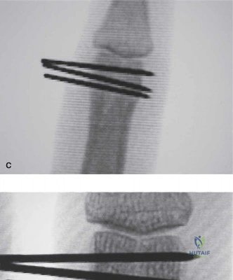
Treating proximal phalanx fractures aims for stable anatomical reduction, joint congruity, and early mobilization. Decision-making considers fracture stability, displacement, articular involvement, and rotational deformity. Operative indications include unstable, irreducible, open, or significantly displaced articular fractures. Stable, minimally displaced non-articular fractures may be managed non-operatively, always prioritizing functional recovery and preventing post-traumatic stiffness.
A 28-year-old amateur rugby player presents with a closed, isolated proximal phalanx shaft fracture of the middle finger following a direct axial load injury. Clinical examination shows mild swelling, and the patient reports pain on movement. You have obtained the following imaging. How would you describe the fracture morphology, and what are your primary clinical concerns regarding the decision for operative versus non-operative management?
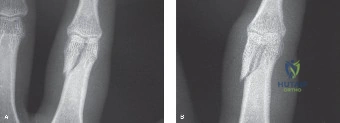
Candidate: I would describe the fracture as a displaced, angulated, and potentially shortened fracture of the proximal phalanx. My main concern is that any malalignment will cause scissoring of the fingers. I would look at the radiographs to check for angulation, shortening, and rotation, and then decide if it needs a splint or surgery.
The candidate fails to use precise orthopaedic terminology (e.g., apex volar/dorsal angulation) and ignores the clinical assessment of rotation. They also fail to mention the "digital cascade," which is the critical clinical check for rotational malalignment.
The candidate must systematically evaluate: 1. Fracture pattern (e.g., transverse vs. oblique). 2. Deformation: Specify the apex direction (e.g., proximal shaft fractures typically show apex volar angulation due to intrinsic pull). 3. Clinical Thresholds: State the "rules of thumb"—less than 10 degrees angulation, <2mm shortening, and zero rotational deformity for non-operative care. 4. Assessment: Emphasize that clinical assessment of the digital cascade during active flexion is superior to radiographs for detecting rotational malalignment.
You have decided to proceed with open reduction and internal fixation for this proximal phalanx fracture. As you plan the approach, discuss the trade-offs between the dorsal approach and the midaxial approach. Which one do you prefer and why?
Candidate: A dorsal approach is easier because it's a direct route to the bone, but it risks damaging the extensor tendons. A midaxial approach is harder because you have to protect the neurovascular bundle, but it’s better for the tendons. I would use the dorsal approach for a simple fracture.
Missing the mention of the "Champeau" or "tendon-sparing" interval in the dorsal approach, and failing to acknowledge that dorsal plating itself causes the adhesions, not just the approach.
The candidate should structure the answer by anatomy: 1. Dorsal Approach: Acknowledge the tendon-splitting vs. tendon-sparing (interval) techniques. Mention that dorsal hardware prominence is a known cause of adhesions. 2. Midaxial Approach: Highlight its use in the "functionally silent zone" between Grayson’s and Cleland’s ligaments, protecting the neurovascular bundle. 3. Preference: Choose based on fracture comminution—lateral plating (via midaxial) is preferred to minimize extensor tendon interference, whereas dorsal plating is reserved for specific tension band needs.
Post-operatively, the patient has developed significant stiffness at the proximal interphalangeal (PIP) joint. Describe your management strategy for this complication.
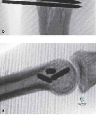
Candidate: I would send the patient to hand therapy for aggressive stretching. If that fails after a couple of months, I would consider surgical tenolysis or a capsulotomy to release the tight joint.
Failing to mention the absolute requirement of radiographic union before attempting tenolysis, and failing to define the timeline (3–6 months) before considering surgery.
The candidate must outline a staged protocol: 1. Non-operative: Intensive hand therapy (active/passive motion, dynamic splinting). 2. Prerequisite for Surgery: Ensure solid radiographic union—tenolysis on a non-healed fracture is catastrophic. 3. Surgical Management: Describe the release of adhesions around the extensor/flexor mechanisms and, if necessary, capsulotomy (dorsal/volar). 4. Post-op: Emphasize immediate active motion, possibly using indwelling regional analgesia to facilitate pain-free rehab.
