Arthroscopic Débridement: Solving Elbow Degenerative Joint Disease Pain
Key Takeaway
Here are the crucial details you must know about Arthroscopic Débridement: Solving Elbow Degenerative Joint Disease Pain. Elbow degenerative joint disease, or primary degenerative arthritis of the elbow, is a relatively rare condition. It involves the loss and fragmentation of cartilage, leading to loose bodies, osteophyte formation, and progressive joint contractures. Commonly affecting manual laborers and athletes, it causes symptoms like pain, loss of extension, and mechanical issues such as catching or locking.
Introduction and Epidemiology
Primary degenerative joint disease of the elbow is a relatively uncommon clinical entity when compared to osteoarthritis of weight-bearing joints such as the hip or knee. The typical demographic profile consists of middle-aged males with a history of heavy manual labor, repetitive upper extremity weight-bearing, or overhead athletic activities. Weightlifters, construction workers, and individuals who rely heavily on their upper extremities for ambulation, such as wheelchair users or those dependent on crutches, represent the most frequently affected patient populations.
Unlike inflammatory arthropathies, which typically present with diffuse cartilage destruction and global joint space narrowing, primary elbow osteoarthritis is characterized by hypertrophic osteophyte formation, capsular contracture, and loose body generation, often with relative preservation of the articular cartilage in the mid-arc of the ulnohumeral joint. The pathogenesis involves a triad of cartilage fragmentation leading to loose bodies, reactive osteophyte formation at the articular margins, and progressive thickening and contracture of the joint capsule.

Patients typically present with a chief complaint of progressive loss of motion and pain at the extremes of flexion and extension. Mid-arc motion is frequently painless, which is a critical diagnostic feature distinguishing primary degenerative joint disease from inflammatory or advanced post-traumatic arthritis. Mechanical symptoms such as catching, locking, and painful crepitus are common and correlate with the presence of intra-articular loose bodies or prominent osteophytes. Concomitant ulnar neuropathy at the cubital tunnel is frequently observed, driven by traction from valgus deformity, direct compression from medial osteophytes, or chronic joint effusion.
While total elbow arthroplasty provides excellent pain relief for low-demand patients with inflammatory arthritis, its application in the young, active, or heavy-laboring demographic is contraindicated due to an unacceptably high rate of early aseptic loosening and catastrophic failure. Elbow arthrodesis, while durable, results in severe functional limitation and is rarely accepted by patients. Consequently, joint-preserving procedures such as arthroscopic débridement, osteophyte resection, and capsulectomy have emerged as the gold standard for managing symptomatic primary elbow osteoarthritis in the active patient.
Surgical Anatomy and Biomechanics
A profound understanding of elbow osseous anatomy, capsuloligamentous structures, and the proximity of major neurovascular bundles is paramount for safe and effective arthroscopic intervention. The elbow is a highly congruent, complex hinge joint comprising the ulnohumeral, radiocapitellar, and proximal radioulnar articulations. The primary constraints to motion in the degenerative elbow are bony impingement and capsular contracture.
Anteriorly, the coronoid process of the ulna articulates with the coronoid fossa of the distal humerus during terminal flexion. In the degenerative state, hypertrophic osteophytes develop on the tip of the coronoid and within the coronoid fossa, resulting in mechanical block to flexion. Similarly, the radial head articulates with the radial fossa of the humerus, where osteophytes can further restrict terminal flexion and forearm rotation.

Posteriorly, the olecranon process seats perfectly within the olecranon fossa of the distal humerus during terminal extension. Osteophyte formation at the tip of the olecranon and within the olecranon fossa creates a direct mechanical block to extension. This bony impingement is frequently compounded by the presence of multiple cartilaginous or osseous loose bodies residing within the posterior compartment or the olecranon fossa itself.
The joint capsule in the osteoarthritic elbow undergoes significant pathologic changes. The anterior capsule becomes fibrotic, thickened, and contracted, tethering the joint and restricting extension. The posterior capsule undergoes similar fibrotic changes, restricting flexion.
Neurovascular anatomy dictates portal placement and the limits of safe resection. The median nerve and brachial artery lie immediately anterior to the brachialis muscle. The radial nerve traverses the anterolateral aspect of the joint, crossing the radiocapitellar joint line. The ulnar nerve resides in the cubital tunnel posteromedially, directly posterior to the medial epicondyle. Intimate knowledge of these structures is critical, particularly during anterior capsulectomy and when establishing the proximal anteromedial and anterolateral portals.

Indications and Contraindications
Patient selection is the primary determinant of success in arthroscopic management of elbow degenerative joint disease. The ideal candidate has preserved mid-arc articular cartilage but suffers from mechanical blocks to motion and terminal pain due to osteophytes and capsular contracture.
| Clinical Scenario | Operative Indication | Non Operative Indication |
|---|---|---|
| Mild Osteoarthritis | Failed conservative management > 6 months | First-line therapy NSAIDs activity modification |
| Mechanical Symptoms | Catching locking loose bodies present | Asymptomatic incidental findings on imaging |
| Motion Loss | Functional deficit impacting ADLs or work | Mild deficit not impacting daily function |
| Pain Characteristics | Pain strictly at extremes of motion terminal pain | Severe mid-arc pain diffuse cartilage loss |
| Ulnar Neuropathy | Progressive motor sensory deficit | Mild intermittent symptoms manageable with splinting |
| Joint Stability | Stable joint architecture | Gross instability requiring complex reconstruction |
Contraindications to arthroscopic débridement include active intra-articular infection, severe architectural distortion of the joint that precludes safe portal placement, and advanced global osteoarthritis with severe mid-arc pain. In patients with complete loss of joint space and bone-on-bone articulation throughout the entire arc of motion, arthroscopic débridement is unlikely to provide durable pain relief, although it may still be utilized strictly for the removal of loose bodies if mechanical locking is the primary complaint. Prior ulnar nerve transposition requires meticulous attention, as the nerve may be vulnerable during the establishment of medial portals.
Pre Operative Planning and Patient Positioning
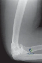
Comprehensive preoperative evaluation relies on a combination of high-quality imaging and meticulous physical examination. Standard anteroposterior, lateral, and oblique radiographs of the elbow are mandatory to assess joint space narrowing, identify loose bodies, and evaluate the extent of osteophyte formation.
Advanced imaging, specifically non-contrast computed tomography with three-dimensional surface rendering, is highly recommended. CT provides exceptional detail regarding the location and volume of osteophytes within the coronoid and olecranon fossae, the exact morphology of the olecranon tip, and the precise location of intra-articular loose bodies. Magnetic resonance imaging is generally less useful for surgical templating of bony resections but may be indicated if concomitant ligamentous pathology or osteochondritis dissecans is suspected.

Patient positioning is a critical component of the procedure. The lateral decubitus position is favored by many high-volume elbow arthroscopists. The patient is placed in the lateral position with the operative arm supported over a padded post or suspended via an arm holder. This position allows excellent access to both the anterior and posterior compartments without the need to reposition the patient intraoperatively. The forearm is allowed to hang freely, utilizing gravity to apply a gentle distraction force across the joint.
Alternatively, the prone position can be utilized, allowing the arm to hang over the side of the operating table. The supine suspended position is another viable option, particularly for surgeons who prefer the arm suspended overhead with traction. Regardless of the position chosen, a sterile tourniquet is typically applied high on the brachium to ensure adequate visualization, although it should be deflated periodically if the procedure is prolonged to minimize ischemic risk.
Fluid management is of paramount importance. Gravity inflow or a mechanical pump set to low pressure (typically 30-40 mmHg) should be used to minimize fluid extravasation into the surrounding soft tissues, which can rapidly lead to compartment syndrome of the forearm or arm.
Detailed Surgical Approach and Technique
The surgical technique for arthroscopic débridement of the elbow requires a systematic, compartmentalized approach to ensure complete pathology addressment while minimizing neurovascular risk.
Portal Placement and Joint Distension
The procedure begins with joint distension. Approximately 15 to 20 mL of sterile normal saline is injected into the joint through the soft spot (the center of the triangle formed by the lateral epicondyle, radial head, and olecranon tip) to displace the neurovascular structures anteriorly and expand the capsular volume.
The proximal anteromedial portal is typically established first. The entry point is 2 cm proximal and 1 cm anterior to the medial epicondyle. A scalpel is used to incise the skin only, and a blunt trocar is directed toward the center of the joint, maintaining contact with the anterior humerus to avoid the median nerve and brachial artery.
Once intra-articular placement is confirmed, diagnostic arthroscopy of the anterior compartment is performed. Under direct visualization, the proximal anterolateral portal is established. This portal is located 2 cm proximal and 1 cm anterior to the lateral epicondyle. An 18-gauge spinal needle is used to localize the ideal trajectory, aiming for the center of the joint anterior to the radiocapitellar articulation. A skin incision is made, and a blunt trocar is advanced to avoid injury to the radial nerve.
Anterior Compartment Debridement
With both anterior portals established, the surgeon can alternate viewing and working portals to address anterior pathology comprehensively. The initial focus is the removal of loose bodies and the synovectomy required to visualize the bony architecture.
Attention is directed to the coronoid fossa and the radial fossa. A motorized shaver and an arthroscopic burr are utilized to resect hypertrophic synovium and subsequently remove osteophytes. The goal is to restore the normal depth and contour of the coronoid fossa to accommodate the coronoid process during terminal flexion.

Osteophytes on the tip of the coronoid process and the radial head are also resected. Care must be taken during radial head débridement to avoid iatrogenic injury to the lateral ulnar collateral ligament.
If a flexion contracture is present, an anterior capsulectomy is performed. Using an arthroscopic basket punch or a radiofrequency ablation wand, the anterior capsule is resected from medial to lateral. The resection must stay strictly on the capsular tissue, avoiding penetration into the brachialis muscle, which serves as the primary barrier protecting the median and radial nerves. The capsulectomy typically begins proximally at the level of the joint line and proceeds distally.
Posterior Compartment Debridement
Following completion of the anterior work, attention is shifted to the posterior compartment. The direct posterior portal (3 cm proximal to the olecranon tip in the midline) and the posterolateral portal (just lateral to the triceps tendon) are established.
The posterior compartment is frequently the site of numerous loose bodies and extensive osteophyte formation. The olecranon fossa is systematically cleared of fibrotic tissue and loose bodies. The hypertrophic osteophytes on the medial and lateral margins of the olecranon fossa are resected using an arthroscopic burr.
The tip of the olecranon is a primary source of impingement in extension. An aggressive resection of the olecranon tip osteophytes is performed. The burr is used to contour the olecranon until it seats deeply within the olecranon fossa without impingement during full extension.
In cases of advanced impingement, an arthroscopic Outerbridge-Kashiwagi procedure (transhumeral fenestration) may be performed. This involves utilizing a burr to create a fenestration through the olecranon fossa into the coronoid fossa. This technique effectively deepens both fossae, accommodates residual osteophytes, and allows access to the anterior compartment from the posterior portals.
If an extension contracture persists after bony resection, a posterior capsulectomy is performed. The posterior capsule is resected from the medial to the lateral gutter, taking care to protect the ulnar nerve medially.
Complications and Management
Arthroscopic management of elbow degenerative joint disease is a technically demanding procedure associated with a unique set of potential complications. The most devastating complications involve iatrogenic injury to the major neurovascular structures.

Nerve injuries can occur during portal placement, excessive retraction, or aggressive capsular resection. The ulnar nerve is particularly vulnerable during posterior capsulectomy and débridement of the medial gutter. The radial nerve is at risk during anterolateral portal establishment and lateral capsulectomy. The median nerve is at risk if the anterior capsule is breached and instruments enter the brachialis muscle.
Fluid extravasation is a constant threat during elbow arthroscopy. The fascial compartments of the arm and forearm have limited compliance. Prolonged surgical times, high pump pressures, and extensive capsulotomies increase the risk of massive fluid extravasation, which can rapidly progress to acute compartment syndrome.
Heterotopic ossification is another recognized complication, particularly following extensive osteophyte resection and capsulectomy. Prophylaxis with nonsteroidal anti-inflammatory drugs (NSAIDs) or localized radiation therapy may be considered in high-risk patients.
| Complication | Estimated Incidence | Prevention and Salvage Strategy |
|---|---|---|
| Transient Neuropraxia | 2% to 10% | Avoid over-distension limit tourniquet time observation and EMG at 6 weeks if no recovery |
| Major Nerve Transection | < 1% | Strict adherence to portal anatomy stay on capsule during resection immediate open exploration and microsurgical repair |
| Compartment Syndrome | < 1% | Low pressure fluid management frequent palpation of forearm immediate fasciotomy if diagnosed |
| Heterotopic Ossification | 3% to 5% | Meticulous hemostasis post-operative NSAIDs (Indomethacin) surgical excision after maturation if symptomatic |
| Infection | 1% to 2% | Preoperative antibiotics sterile technique arthroscopic irrigation and débridement with targeted antibiotics |
| Persistent Stiffness | 5% to 15% | Aggressive postoperative rehabilitation static progressive splinting revision arthroscopic or open release |
Post Operative Rehabilitation Protocols
The success of arthroscopic débridement for elbow osteoarthritis is intrinsically linked to the postoperative rehabilitation protocol. The surgical intervention provides the mechanical capacity for motion, but the rehabilitation process secures and maintains that motion.
Immediate postoperative mobilization is critical. The operative extremity is placed in a bulky soft dressing, avoiding rigid immobilization. Active and active-assisted range of motion exercises are initiated on postoperative day one.
During the acute phase (0 to 2 weeks), the primary goals are edema control, pain management, and the restoration of terminal extension and flexion. Continuous passive motion (CPM) machines may be utilized, although frequent, dedicated active-assisted stretching sessions are generally preferred. Cryotherapy and elevation are essential to manage postoperative swelling, which can mechanically block motion.
In the intermediate phase (2 to 6 weeks), therapy intensifies. If the patient exhibits a persistent deficit in terminal motion, static progressive splinting or dynamic splinting is initiated. Turnbuckle splints for extension and flexion are highly effective in overcoming residual capsular tightness. Strengthening exercises are generally deferred until full, painless range of motion is achieved to avoid aggravating joint inflammation.
Return to heavy manual labor or overhead athletics is typically permitted between 6 and 12 weeks postoperatively, contingent upon the restoration of functional motion and the resolution of pain. Patients must be counseled that maximal improvement in motion and pain relief may take up to six months to fully manifest.
Summary of Key Literature and Guidelines
The evolution of surgical management for elbow degenerative joint disease has shifted heavily toward arthroscopic techniques over the past two decades. Historically, the open Outerbridge-Kashiwagi (OK) procedure or the open ulnohumeral arthroplasty as described by Morrey were the mainstays of treatment. While these open procedures provided reliable pain relief and improved motion, they were associated with significant morbidity, including triceps weakness, prolonged rehabilitation, and higher risks of incisional complications.

Contemporary literature robustly supports the efficacy and safety of arthroscopic débridement. High-quality retrospective series and comparative cohort studies have consistently demonstrated that arthroscopic management yields equivalent, if not superior, improvements in range of motion and pain scores compared to open techniques, with the added benefits of decreased perioperative morbidity, accelerated rehabilitation, and lower complication rates.
Studies by Savoie, O'Driscoll, and others have validated the durability of arthroscopic osteocapsular arthroplasty. Long-term follow-up indicates that the majority of patients maintain their improvements in motion and experience sustained pain relief for 5 to 10 years postoperatively. While arthroscopic débridement does not halt the underlying degenerative process, it effectively resets the mechanical clock of the joint, significantly delaying the need for total elbow arthroplasty in the younger, high-demand patient population.
Current academic guidelines recommend arthroscopic débridement as the primary surgical intervention for symptomatic primary elbow osteoarthritis characterized by mechanical impingement and preserved mid-arc cartilage. The procedure is technically demanding and should be performed by surgeons with advanced training in elbow arthroscopy to minimize the inherent neurovascular risks and maximize functional outcomes. Thorough preoperative templating, meticulous surgical technique, and aggressive postoperative rehabilitation remain the cornerstones of successful management.
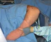
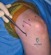
Clinical & Radiographic Imaging