Operative Management of Elbow Contractures and Medial Epicondylitis
Key Takeaway
Elbow contractures significantly impair upper extremity function. While a normal elbow arc is 0 to 150 degrees, a functional range requires 30 to 130 degrees. Management begins with static progressive splinting, advancing to arthroscopic or open arthrolysis for refractory cases. This guide details the pathophysiology of elbow stiffness, risk factors for heterotopic ossification, and the step-by-step Nirschl surgical technique for medial epicondylitis.
Comprehensive Introduction and Patho-Epidemiology
The elbow joint, a highly constrained and complex diarthrodial hinge, is uniquely susceptible to devastating post-traumatic and post-surgical stiffness, as well as chronic degenerative tendinopathies. The operative management of elbow contractures and medial epicondylitis represents a formidable challenge for the orthopedic surgeon, requiring a profound understanding of periarticular soft-tissue envelopes, intricate neurovascular anatomy, and the fundamental biomechanics of upper extremity function. Elbow contractures, defined by a clinically significant reduction in the normal functional arc of motion, arise from a diverse array of intrinsic and extrinsic etiologies. The pathophysiology of post-traumatic elbow stiffness is driven by an aggressive fibroproliferative cascade. Following trauma, local hypoxia and the release of inflammatory cytokines, particularly Transforming Growth Factor-beta 1 (TGF-β1), stimulate the differentiation of local fibroblasts into myofibroblasts. These cells, rich in alpha-smooth muscle actin, orchestrate the deposition of disorganized type I and type III collagen, leading to a thickened, non-compliant joint capsule. Furthermore, the elbow is notoriously prone to heterotopic ossification (HO), an ectopic bone formation process driven by osteoprogenitor cells within the periarticular soft tissues responding to neurogenic, thermal, or severe mechanical trauma.
Concurrently, medial epicondylitis, classically termed "golfer's elbow," represents a distinct pathophysiological entity affecting the common flexor-pronator origin at the medial epicondyle. Despite the historical nomenclature implying an acute inflammatory "-itis," exhaustive histological analyses have definitively characterized the lesion as a chronic, non-inflammatory, degenerative tendinopathy. The hallmark histopathological finding is angiofibroblastic hyperplasia, a process characterized by mucinoid degeneration, disorganized collagen fiber architecture, and an abortive neovascularization response. This degenerative cascade is typically precipitated by repetitive microtrauma and eccentric loading of the pronator teres (PT) and the flexor carpi radialis (FCR). Over time, the structural integrity of the tendon microarchitecture fails, leading to microtearing, chronic pain, and profound functional impairment in gripping and forearm pronation activities.
Epidemiologically, elbow contractures are a ubiquitous complication following severe elbow trauma, particularly terrible triad injuries, complex distal humerus fractures, and prolonged periods of rigid immobilization. The incidence of clinically significant stiffness following operative fixation of elbow trauma can approach 15% to 20%, despite modern advances in rigid internal fixation and early mobilization protocols. Heterotopic ossification is particularly prevalent in patients sustaining concurrent traumatic brain injuries (TBI) or extensive thermal burns, where the incidence of massive, bridging ectopic bone can exceed 30%. Conversely, medial epicondylitis is less common than its lateral counterpart (tennis elbow), accounting for approximately 10% to 20% of all epicondylitis cases. It predominantly affects individuals in their fourth to sixth decades of life, with a higher prevalence in laborers engaged in repetitive forceful gripping, overhead throwing athletes, and, classically, golfers.
The intersection of these two pathologies often manifests in the complex elbow patient. A patient presenting with post-traumatic stiffness may also harbor underlying tendinopathic changes, or the surgical approaches required to address massive medial heterotopic ossification may inadvertently compromise the flexor-pronator mass. Therefore, a comprehensive mastery of both the arthrolytic techniques required to liberate the stiff elbow and the precise, tissue-sparing excisional techniques required to eradicate angiofibroblastic hyperplasia is essential for the advanced upper extremity surgeon. The primary objective in managing these conditions is the restoration of a functional, painless arc of motion, allowing the patient to seamlessly perform activities of daily living without biomechanical compromise.
Detailed Surgical Anatomy and Biomechanics
A rigorous comprehension of elbow surgical anatomy and biomechanics is the cornerstone of successful operative intervention. The elbow is a highly congruent trochginglymoid joint, comprising three distinct articulations: the ulnohumeral, radiocapitellar, and proximal radioulnar joints. The normal anatomical range of motion is 0 to 150 degrees of flexion, with 75 degrees of pronation and 85 degrees of supination. However, extensive biomechanical investigations pioneered by Morrey and colleagues have established that the functional arc of motion—the range required to perform the vast majority of activities of daily living (ADLs)—spans from 30 to 130 degrees of flexion, coupled with 50 degrees of pronation and 50 degrees of supination. The primary stabilizers of the elbow include the highly congruent ulnohumeral articulation, the medial collateral ligament (MCL) complex, and the lateral collateral ligament (LCL) complex. The anterior bundle of the MCL (AMCL) is the primary restraint to valgus stress, originating from the anteroinferior aspect of the medial epicondyle and inserting onto the sublime tubercle of the proximal ulna.
The joint capsule itself plays a critical role in the pathogenesis of elbow stiffness. The anterior capsule is relatively thin but broad, spanning from the radial and coronoid fossae proximally to the annular ligament and coronoid process distally. In the setting of a flexion contracture, the anterior capsule becomes profoundly thickened, contracted, and adherent to the underlying brachialis muscle. The posterior capsule, which attaches proximal to the olecranon fossa and distally to the olecranon, similarly undergoes fibrotic shortening in extension contractures. The intimate relationship between the anterior capsule and the neurovascular structures of the antecubital fossa dictates that anterior capsulectomy must be performed with meticulous dissection, often requiring the brachialis to be elevated off the capsule to serve as a protective barrier for the brachial artery and median nerve.
The precise anatomy of the medial epicondyle and the common flexor origin is paramount when addressing medial epicondylitis. The flexor-pronator mass comprises, from proximal to distal and anterior to posterior, the pronator teres (PT), flexor carpi radialis (FCR), palmaris longus (PL), flexor digitorum superficialis (FDS), and flexor carpi ulnaris (FCU). The pathological lesion in medial epicondylitis is most frequently localized to the deep interface between the origins of the PT and the FCR. Crucially, the AMCL lies immediately deep to the flexor-pronator mass. The surgeon must possess an acute spatial awareness to resect the overlying angiofibroblastic tissue without violating the underlying AMCL, an iatrogenic error that precipitates devastating valgus instability.
Furthermore, the cutaneous and major peripheral nerve anatomy dictates the surgical approaches to both the stiff elbow and medial epicondylitis. The medial antebrachial cutaneous (MABC) nerve represents a critical anatomical hazard during medial approaches. The MABC typically arborizes 2 to 3 centimeters proximal to the medial epicondyle, sending sensory branches that cross anterior and distal to the epicondyle. Inadvertent transection or entrapment of these branches during a Nirschl procedure or a medial column release results in a highly morbid, recalcitrant neuroma. Similarly, the ulnar nerve, coursing through the cubital tunnel just posterior to the medial epicondyle, is intimately associated with the posterior band of the MCL and the medial capsule. In cases of severe contracture, particularly those involving medial heterotopic ossification, the ulnar nerve is frequently encased in dense scar or bone, necessitating meticulous neurolysis and often anterior transposition prior to any medial arthrolytic maneuvers.
Exhaustive Indications and Contraindications
The decision to proceed with operative intervention for elbow contractures or medial epicondylitis must be predicated on a rigorous evaluation of the patient's functional deficits, the failure of exhaustive non-operative modalities, and a careful assessment of the biological and mechanical state of the joint. For elbow contractures, the initial management is universally non-operative, relying on the viscoelastic properties of the joint capsule. A stringent, 3- to 6-month regimen of static progressive or dynamic splinting, utilizing the principle of stress relaxation, is mandatory. Surgical arthrolysis is indicated only when a patient demonstrates a persistent functional deficit (typically a flexion contracture exceeding 30-45 degrees or flexion less than 130 degrees) that has plateaued despite absolute compliance with a maximal non-operative rehabilitation program. In cases of heterotopic ossification, surgical excision is indicated once the ectopic bone has biologically matured, typically evidenced by sharp, well-defined trabecular markings on plain radiographs and a normalized serum alkaline phosphatase level, minimizing the risk of catastrophic recurrence.
For medial epicondylitis, the indications for surgical intervention are similarly stringent. The vast majority of patients will achieve symptomatic resolution with a comprehensive non-operative algorithm encompassing activity modification, counterforce bracing, eccentric strengthening protocols, and orthobiologic interventions such as Platelet-Rich Plasma (PRP) injections. Operative intervention, typically via the Nirschl procedure, is strictly reserved for patients with recalcitrant, functionally limiting medial elbow pain that has persisted for a minimum of 6 to 12 months despite exhaustive conservative management. The patient must demonstrate localized tenderness directly over the PT/FCR origin, exacerbated by resisted forearm pronation and wrist flexion.
Contraindications must be meticulously respected to avoid catastrophic outcomes. Active intra-articular or periarticular infection is an absolute contraindication to any elective arthrolysis or tendon debridement. In the setting of elbow contractures, advanced articular destruction (e.g., severe post-traumatic osteoarthritis, inflammatory arthropathy with joint space obliteration) represents a relative contraindication to isolated arthrolysis; these patients are more appropriately managed with interposition or total elbow arthroplasty. Furthermore, a non-compliant patient who is unwilling or unable to participate in the grueling, mandatory post-operative rehabilitation protocol is an absolute contraindication for contracture release, as the joint will inevitably re-stiffen. For medial epicondylitis, isolated ulnar neuritis without concomitant flexor-pronator tendinopathy is a contraindication to the Nirschl procedure; these patients require isolated cubital tunnel release or transposition.
| Pathology / Procedure | Primary Indications | Absolute Contraindications | Relative Contraindications |
|---|---|---|---|
| Elbow Arthrolysis (Contracture) | Functional ROM deficit (arc < 100°); Failure of 3-6 months static progressive splinting; Mature Heterotopic Ossification causing mechanical block. | Active joint infection; Patient inability to comply with rigorous post-op CPM/PT; Active, immature HO (high risk of recurrence). | Severe articular cartilage destruction (consider arthroplasty); Severe complex regional pain syndrome (CRPS). |
| Nirschl Procedure (Medial Epicondylitis) | Refractory pain > 6-12 months; Failure of PT, bracing, and PRP/steroid injections; MRI evidence of high-grade tendinosis/partial tearing of PT/FCR. | Cervical radiculopathy (C6/C7) masquerading as elbow pain; Isolated ulnar neuritis; Active local skin infection. | Concomitant profound MCL insufficiency (requires concurrent ligamentous reconstruction); Unmanaged psychiatric comorbidities affecting pain perception. |
Pre-Operative Planning, Templating, and Patient Positioning
Meticulous pre-operative planning is the sine qua non of successful elbow surgery. For the contracted elbow, advanced imaging is mandatory. While standard orthogonal radiographs provide baseline information regarding joint congruity, osteophytes, and the presence of heterotopic ossification, a fine-cut computed tomography (CT) scan with 3-dimensional reconstructions is indispensable. The 3D CT scan allows the surgeon to precisely map the location and volume of osteophytes within the olecranon and coronoid fossae, and to delineate the exact anatomical relationship of heterotopic bone bridges to critical neurovascular structures. If the structural integrity of the collateral ligaments is in question, particularly following prior trauma or multiple surgeries, a magnetic resonance imaging (MRI) arthrogram should be obtained to evaluate the MCL and LCL complexes.
For medial epicondylitis, while the diagnosis is primarily clinical, an MRI without contrast (using fluid-sensitive T2 or STIR sequences) is highly recommended for surgical candidates. The MRI serves a dual purpose: it confirms the presence of tendinosis, characterized by increased signal intensity and thickening within the common flexor origin, and it critically evaluates the integrity of the underlying anterior bundle of the MCL. Pre-operative identification of concurrent MCL insufficiency alters the surgical plan entirely, necessitating a combined debridement and ligamentous reconstruction. Furthermore, electromyography and nerve conduction velocities (EMG/NCV) should be obtained in any patient exhibiting signs of ulnar neuropathy to objectively quantify the degree of cubital tunnel syndrome, dictating the need for a concurrent ulnar nerve transposition.
Patient positioning and anesthesia require careful coordination between the surgical and anesthesia teams. A regional anesthetic, typically an ultrasound-guided supraclavicular or axillary brachial plexus block, is highly preferred. This provides excellent intra-operative muscle relaxation, minimizing the force required for retraction, and offers profound post-operative analgesia, which is critical for initiating immediate continuous passive motion (CPM) therapy. General anesthesia is often utilized as an adjunct to ensure patient comfort during prolonged arthrolytic procedures.
The patient is positioned supine on the operating table. For open arthrolysis and medial epicondylitis procedures, the operative arm is extended onto a radiolucent hand table. A sterile pneumatic tourniquet is applied high on the brachium. The arm is prepped and draped freely to allow for full, unrestricted assessment of the flexion-extension and pronation-supination arcs throughout the procedure. If an arthroscopic arthrolysis is planned, the surgeon may opt for the lateral decubitus position with the arm suspended over a bolster, or the prone position, depending on surgeon preference and training. Regardless of the approach, pre-operative templating must include the availability of a hinged external fixator system. The surgeon must be prepared to apply the fixator if the sequential release of the contracted capsuloligamentous structures results in iatrogenic joint instability, requiring precise identification of the axis of rotation at the center of the capitellum and the anteroinferior medial epicondyle.
Step-by-Step Surgical Approach and Fixation Technique
Open Elbow Arthrolysis: The Column Approach
The open management of severe elbow contractures is best conceptualized through the sequential column approach popularized by Morrey. This technique allows for the systematic release of contracted structures while meticulously preserving the primary ligamentous stabilizers.
- Lateral Column Release: A lateral incision is made, utilizing the Kocher interval (between the anconeus and extensor carpi ulnaris) or the Kaplan interval (between the extensor carpi radialis brevis and extensor digitorum communis). The extensor origin is elevated anteriorly to expose the anterior radiocapitellar joint. The contracted anterior capsule is identified. Using a periosteal elevator, the brachialis muscle is meticulously elevated off the anterior capsule. This is a critical step, as the brachialis serves to protect the brachial artery and median nerve. A complete anterior capsulectomy is performed from lateral to medial.
- Posterior Compartment Clearance: Attention is directed to the posterior aspect of the lateral column. The triceps is elevated off the posterior humerus. The posterior capsule is excised, and the olecranon fossa is cleared of all fibrotic tissue and osteophytes using a high-speed burr or rongeurs. The tip of the olecranon may be resected if it impinges within the fossa during terminal extension.
- Medial Column Release and Ulnar Nerve Management: If a medial contracture or massive medial HO is present, a separate medial incision is utilized. The ulnar nerve must be identified proximally, meticulously neurolysed, and protected. In the setting of severe stiffness, an anterior submuscular or subcutaneous transposition is routinely performed to prevent traction neuritis as motion is restored. The flexor-pronator mass is elevated to expose the medial aspect of the anterior capsule, completing the capsulectomy. The medial collateral ligament must be visualized and rigorously protected throughout this exposure.
- Application of Hinged External Fixation: If the extensive soft-tissue release compromises the LCL or MCL, or if joint distraction is required, a hinged external fixator is applied. The anatomical axis of rotation is identified using fluoroscopy (a perfect lateral view demonstrating concentric circles of the capitellum and trochlear sulcus). A 2.0mm K-wire is driven precisely through the isometric axis. The humeral and ulnar pins are placed, and the hinge is secured over the axis wire, allowing for stable, guided concentric motion.
The Nirschl Procedure for Medial Epicondylitis
The surgical eradication of angiofibroblastic hyperplasia in medial epicondylitis demands precise, tissue-sparing technique to avoid catastrophic complications.
- Incision and MABC Protection: A 5-cm longitudinal incision is made centered slightly posterior to the medial epicondyle. This posterior bias is the most critical step in the approach, designed explicitly to avoid the anteriorly arborizing branches of the Medial Antebrachial Cutaneous (MABC) nerve. Dissection is carried down through the subcutaneous tissue, maintaining meticulous hemostasis.
- Identification of the Pathological Tissue: The common flexor origin is exposed. A longitudinal incision is made directly in line with the fibers of the pronator teres and flexor carpi radialis, beginning at the epicondylar tip and extending distally. The healthy, glistening superficial fibers are retracted to reveal the deeper pathological tissue. The angiofibroblastic tissue is visually distinct: it appears dull, grey, friable, and edematous, entirely lacking the striated, organized appearance of healthy tendon collagen.
- Excision and MCL Preservation: The pathological tissue is excised in an elliptical fashion. The surgeon must exercise extreme caution at the deep margin of the resection. The anterior bundle of the medial collateral ligament (AMCL) lies immediately deep to this tissue. The AMCL must be visually identified and preserved; violation of the AMCL will result in profound iatrogenic valgus instability.
- Epicondylar Decortication and Closure: Once the degenerative tissue is excised, the cortical footprint of the medial epicondyle is lightly decorticated using a 2.0-mm drill bit or a rongeur. This intentional trauma stimulates a localized bleeding response, introducing marrow-derived stem cells and osteoinductive growth factors into the surgical bed to promote robust biological healing. The elliptical defect in the tendon is then closed side-to-side using heavy absorbable sutures (e.g., #1 Vicryl) in an interrupted or figure-of-eight fashion. The skin is closed with a running subcuticular suture.
Complications, Incidence Rates, and Salvage Management
The operative management of the elbow is fraught with potential complications, largely due to the unforgiving nature of the periarticular soft tissues and the dense concentration of critical neurovascular structures. The surgeon must be intimately familiar with the incidence, prevention, and salvage management of these adverse events.
In the treatment of elbow contractures, the most frequent complication is recurrent stiffness. Despite flawless surgical technique, up to 20% of patients will lose a portion of their intra-operative gains due to aggressive post-operative fibroplasia or poor compliance with rehabilitation. Heterotopic ossification recurrence is a devastating complication, occurring in 5% to 10% of high-risk patients despite appropriate prophylaxis. If massive HO recurs and bridges the joint, a minimum of 6 to 12 months must elapse to allow for complete biological maturation before a revision excision can be safely attempted. Nerve injuries, particularly to the ulnar nerve, occur in 2% to 5% of open arthrolyses. Traction neuritis is common following the restoration of profound flexion; therefore, prophylactic ulnar nerve transposition is heavily advocated. Iatrogenic instability resulting from overzealous capsuloligamentous release requires immediate recognition and either primary ligamentous repair or the application of a neutralizing hinged external fixator.
Following the Nirschl procedure for medial epicondylitis, the most feared complication is the formation of a painful neuroma of the Medial Antebrachial Cutaneous (MABC) nerve. Occurring in 1% to 3% of cases, primarily due to improper anterior incision placement, an MABC neuroma is profoundly debilitating. Salvage management requires re-exploration, excision of the neuroma, and burying the proximal nerve stump deep into the belly of the triceps or brachialis muscle to prevent superficial mechanical irritation. Iatrogenic valgus instability, resulting from inadvertent transection of the underlying AMCL during the debridement of the flexor-pronator mass, is a catastrophic error. Patients will present with profound pain and weakness during valgus-loading activities. Salvage necessitates a formal medial collateral ligament reconstruction (Tommy John surgery) utilizing an autograft, typically the palmaris longus or gracilis tendon.
| Complication | Estimated Incidence | Etiology / Risk Factors | Salvage Management / Treatment |
|---|---|---|---|
| Recurrent Stiffness (Arthrolysis) | 15% - 20% | Poor rehab compliance; Inadequate primary release; Aggressive fibroplasia. | Resume static progressive splinting; Manipulation under anesthesia (rarely effective); Revision open arthrolysis. |
| Heterotopic Ossification Recurrence | 5% - 10% | TBI/Burn patients; Premature excision of immature bone; Failure of prophylaxis. | Wait 6-12 months for maturation; Revision excision; Post-op localized radiation (700 cGy) + Indomethacin. |
| MABC Neuroma (Nirschl) | 1% - 3% | Incision placed too far anteriorly; Aggressive superficial retraction. | Surgical exploration, neuroma excision, and deep intramuscular burial of the proximal stump. |
| Iatrogenic Valgus Instability | < 1% | Inadvertent resection of the AMCL during deep tissue debridement. | Formal MCL reconstruction (autograft) using docking or figure-of-eight technique. |
| Ulnar Neuritis | 2% - 5% | Traction from restored flexion; Entrapment in scar tissue; Failure to transpose. | Revision neurolysis and anterior submuscular or subcutaneous transposition. |
Phased Post-Operative Rehabilitation Protocols
The surgical intervention, regardless of its technical perfection, represents merely the first phase of treatment. The ultimate success of both elbow arthrolysis and medial epicondylitis surgery is inextricably linked to a rigorous, phased post-operative rehabilitation protocol.
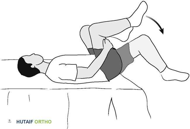
For the patient undergoing elbow arthrolysis, the immediate post-operative goal is the prevention of recurrent capsular adhesions. Phase I (Weeks 0-2) begins in the recovery room. A brachial plexus catheter is often maintained for 48 to 72 hours to provide continuous analgesia, facilitating immediate utilization of a Continuous Passive Motion (CPM) machine. The patient is encouraged to perform active-assisted range of motion (AAROM) exercises out of the CPM multiple times daily. Prophylaxis against heterotopic ossification is initiated immediately, consisting of either a single fraction of localized radiation therapy (700 cGy) within 48 hours, or a 3- to 6-week course of oral Indomethacin (75mg sustained release daily), assuming no gastrointestinal or renal contraindications exist. Phase II (Weeks 2-6) involves the removal of sutures and the initiation of aggressive physical therapy. If the patient exhibits a loss of extension, a static progressive extension splint is fabricated and worn at night. Conversely, flexion deficits are managed with a static progressive flexion splint. Phase III (Weeks 6-12) focuses on terminal stretching and the gradual introduction of isometric strengthening. The patient must be counseled that maximal improvement in range of motion may take up to 12 months to fully realize.
The rehabilitation following a Nirschl procedure for medial epicondylitis is markedly different, prioritizing tendon healing and the gradual re-introduction of load. Phase I: Immediate Postoperative Period (Weeks 0-1) involves immobilization. The arm is placed in a bulky compressive dressing and a rigid posterior plaster splint with the elbow at 90 degrees of flexion and the forearm in neutral rotation. This protects the delicate side-to-side repair of the flexor-pronator mass. Phase II: Early Motion (Weeks 1-3) begins with the removal of the splint. The patient initiates active and active-assisted elbow ROM exercises. Crucially, passive stretching of the wrist flexors and forearm pronators is strictly prohibited during this phase to avoid catastrophic failure of the repair. Phase III: Strengthening (Weeks 3-12) commences only when the patient has achieved a full, painless arc of motion. Rehabilitation transitions to isometric exercises, followed by a highly structured program of concentric and eccentric strengthening of the flexor-pronator musculature. Phase IV: Return to Activity (Months 3+) allows for the resumption of heavy manual labor and sports. Throwing athletes and golfers undergo a specialized, interval return-to-play program, focusing on core stability, kinetic chain mechanics, and gradual increases in velocity and volume to prevent recurrence.
Summary of Landmark Literature and Clinical Guidelines
The contemporary operative management of elbow contractures and medial epicondylitis is heavily informed by decades of rigorous biomechanical research and landmark clinical trials. A profound understanding of this literature is essential for the academic orthopedic surgeon.
The foundational biomechanical parameters of elbow function were established by Morrey, Askew, and Chao in their seminal 1981 publication in the Journal of Bone and Joint Surgery. By utilizing electrogoniometers to evaluate the arc of motion required for 15 diverse activities of daily living, they defined the "functional arc" of the elbow as 30 to 130 degrees of flexion and 50 degrees of both pronation and supination. This single study remains the gold standard metric against which all outcomes of contracture release are measured. The efficacy of non-operative management for elbow stiffness was profoundly validated by Ulrich et al., who demonstrated that the application of static progressive splinting, utilizing the viscoelastic principle of stress relaxation, yielded a mean increase of 26 degrees in the flexion-extension arc, fundamentally shifting the paradigm towards exhaustive conservative management prior to surgical intervention. Furthermore, the classification and management of heterotopic ossification about the elbow are guided by the Hastings and Graham classification system, which delineates the functional limitations imposed by ectopic bone and dictates the timing and approach for surgical excision.
In the realm of medial epicondylitis, the pathological understanding and surgical management were revolutionized by Robert Nirschl. Nirschl and Pettrone's landmark histological studies definitively debunked the concept of an acute inflammatory tendonitis, introducing the term "angiofibroblastic hyperplasia" to describe the chronic, degenerative microtearing of the common flexor origin. The surgical technique bearing his name—the meticulous excision of this pathological tissue with preservation of the underlying MCL—remains the gold standard operative intervention. More recently, clinical guidelines published by the American Academy of Orthopaedic Surgeons (AAOS) and numerous meta-analyses have reinforced the necessity of exhausting non-operative modalities, particularly eccentric exercise protocols and orthobiologic injections (PRP), for a minimum of 6 to 12 months prior to considering surgical debridement for epicondylitis. Mastery of these historical milestones and contemporary guidelines is paramount for delivering evidence-based, state-of-the-art care to the complex elbow patient.
This academic synthesis is based on ---
