Clinical Diagnosis & Imaging of Elbow Adhesive Capsulitis: An Orthopedic Case Study
Key Takeaway
Elbow adhesive capsulitis is diagnosed through a comprehensive clinical evaluation, revealing progressive, symmetrical global motion restriction and a firm capsular end-feel. Predisposing factors like diabetes are considered. Imaging, primarily MRI, confirms capsular thickening and inflammation while ruling out other pathologies, making it crucial for a definitive diagnosis.
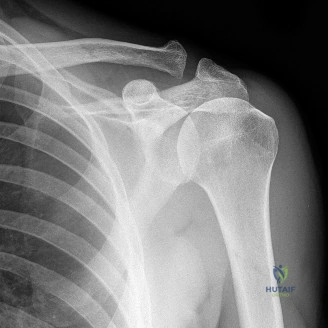
A 48-year-old male presents with 9 months of progressive left elbow pain and stiffness. There is no significant trauma history, but he has Type 2 Diabetes and hypothyroidism. His examination demonstrates a "capsular pattern" of stiffness. Looking at his radiographs provided, how do you categorize this clinical scenario, and what are the primary differential diagnoses you must exclude?
Candidate: Given the patient’s comorbidities of diabetes and thyroid disease, this is likely primary adhesive capsulitis. The radiographs are normal, showing no osteophytes or heterotopic ossification. I would differentiate this from post-traumatic arthrofibrosis, osteoarthritis, and heterotopic ossification (HO).
Candidates often forget to systematically categorize the stiffness. Simply naming the disease is insufficient. Failing to mention why you excluded HO (e.g., lack of radiographic evidence) or ignoring the "capsular pattern" terminology limits the candidate's professional depth.
This is a presentation of primary adhesive capsulitis, categorized as Kay Type I stiffness (soft-tissue contracture without osseous pathology). My differential includes:
1. Post-traumatic arthrofibrosis: Excluded by lack of significant injury and lack of articular incongruity.
2. Elbow OA: Excluded by well-maintained joint spaces and lack of marginal osteophytes.
3. Heterotopic Ossification: Excluded by the absence of calcific densities on radiographs.
The "capsular pattern" (symmetrical flexion/extension loss) with preserved radioulnar rotation is the classic clinical sign of an intrinsic capsular process.
The patient has failed 6 months of non-operative management. You decide to proceed with arthroscopic release. Why is the distinction between "capsulotomy" and "capsulectomy" critical in this patient, and what is your specific strategy for the anterior compartment?
Candidate: A capsulotomy is just an incision, but a capsulectomy is a formal excision of the fibrotic capsule. We need to perform a capsulectomy to prevent recurrence. Anteriorly, I would resect the capsule until I see the brachialis muscle.
Candidates often miss the safety aspect—failing to mention the proximity of the median nerve and brachial artery during the anterior release. They also fail to emphasize why the brachialis is a "target" (acting as a dynamic interpositional graft).
The distinction is vital: capsulotomy carries a high risk of re-adhesion. A capsulectomy is essential to remove the pathological matrix.
Anterior Strategy: Using radiofrequency and a shaver, I perform an extensive release from the lateral gutter to the medial side. The endpoint is the clear visualization of the brachialis muscle fibers. This serves as a vascularized, dynamic interposition layer. Crucially, I must remain aware of the neurovascular bundle (median nerve/brachial artery) located immediately anterior to the capsule, keeping the shaver blade facing the joint at all times.
What is your postoperative protocol to ensure this patient does not revert to his preoperative stiffness?
Candidate: I would use a regional nerve block for pain control, early aggressive physical therapy, and a CPM machine. I might use Indomethacin to prevent HO.
A weak answer fails to mention "static progressive splinting" or the concept of low-load, prolonged stretch. Examiners want to know you understand that early motion is not just "PT," but a structured, biomechanical approach to collagen remodeling.
The success of the procedure relies heavily on the postoperative phase:
1. Immediate Analgesia: Indwelling supraclavicular catheter to allow immediate, pain-free ROM.
2. Continuous Passive Motion (CPM): Used for 18–20 hours/day in the first week to maintain the intraoperative range.
3. Pharmacology: Indomethacin for 3–6 weeks as prophylaxis against HO.
4. Static Progressive Splinting: Transitioning to splints that provide low-load, prolonged stretch, which is biomechanically superior to high-load brief stretching for modifying collagen cross-linking.
5. Patient Education: Clear counseling that the "rehab is as important as the surgery" to manage expectations.
