Comprehensive Case Study: High-Energy Multiple Metacarpal Fractures with Open Injury Suspicion
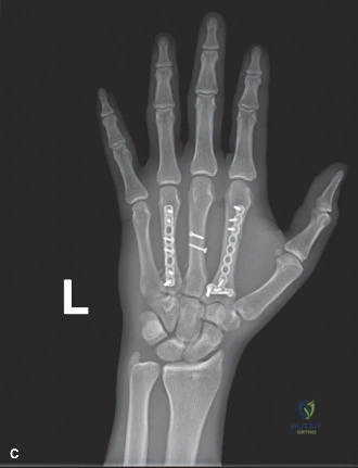
Key Takeaway
High-energy multiple metacarpal fractures, often from crush injuries, present with severe pain, deformity, swelling, and potential open wounds. Clinical examination reveals gross instability, crepitus, and disrupted digital cascade. Diagnosis is confirmed with three-view X-rays showing significant angulation, shortening, and rotational malalignment, crucial for guiding definitive management, especially with suspected open fractures.
A 32-year-old construction worker presents with a high-energy crush injury to the dominant hand. On examination, you note an obvious disruption of the digital cascade, specifically scissoring of the 5th digit over the 4th during gentle flexion. What is the most critical clinical concern here, and what does it indicate about the fracture pattern?
Candidate: The scissoring of the 5th digit indicates rotational malalignment at the fracture site. This is a critical finding because rotational deformities are poorly tolerated by the hand and can lead to significant functional impairment—specifically, the fingers crossing over each other when making a fist. It suggests an unstable fracture pattern, likely a spiral or long oblique metacarpal shaft fracture.
Focusing solely on the dorsal angulation or the bony fracture. Candidates often fail to link the "scissoring" sign specifically to rotational instability and the resulting functional "overlap" of the digits during gripping, which is the key patient-reported outcome concern.
Highlight that rotational malalignment is the most common cause of post-traumatic functional impairment in metacarpal fractures. Emphasize that because the hand is a multi-articulated lever system, even 5-10 degrees of rotation at the metacarpal level translates to a 1.5–2 cm discrepancy at the fingertip. State clearly: "Rotational correction is the primary priority in achieving an anatomical reduction, as it is notoriously difficult to correct once the fracture is united."
You review the initial imaging. Given the multi-metacarpal nature of the injury, describe your radiographic assessment strategy and the specific radiographic signs you are looking for in the 5th metacarpal.
Candidate: I would assess the 2nd through 5th metacarpals for length, rotation, and sagittal alignment. For the 5th metacarpal specifically, I am evaluating the degree of dorsal angulation—which usually has a higher tolerance than the 2nd—and the spiral component which dictates stability. I would use the oblique view to confirm no intra-articular extension into the CMC joint and assess for comminution that would preclude stable fixation.
Failing to mention the "True Lateral" view for assessing dorsal angulation or failing to mention the assessment of the CMC joint. Examiners look for the candidate to recognize that 2nd/3rd metacarpal fractures require near-perfect reduction, whereas 4th/5th tolerate more angulation due to the mobility of the CMC joints.
Structure the answer by the "Stability Principle": 1. Assessment: Measure length relative to the 3rd metacarpal. 2. Tolerance: Contrast the 2nd/3rd (rigid, 10-15 degree tolerance) with the 4th/5th (mobile, 20-30 degree tolerance). 3. Mechanical sign: The "overlap" on the PA view in this case is pathognomonic for rotational instability in a spiral fracture. 4. Joint Integrity: Specifically rule out CMC dislocation, which is a surgical emergency.
The fractures are Gustilo-Anderson Type I. How does this influence your operative planning compared to a closed injury, and what are your priorities for the surgical approach?
Candidate: Even though they are Type I, they are still open fractures. My priorities are: immediate systemic antibiotics, formal debridement of the wound and fracture site under irrigation, and then stable internal fixation. I would avoid excessive periosteal stripping to prevent devascularization, especially given the crush mechanism.
Assuming that because it's a small wound, "simple" debridement is enough. Failing to mention that the anatomy of the hand is unforgiving—stripping the periosteum here significantly elevates the risk of nonunion in a patient who is already a smoker.
The answer must be systematic: 1. Tetanus & Antibiotics: The "Golden Hour" principle for open fractures. 2. Debridement: "Formal" debridement includes formal extension of the lacerations to inspect the fracture site, not just cleaning the skin. 3. Fixation: Use a low-profile locking plate construct to allow early range of motion, which is the best defense against stiffness in a crush-injured hand. 4. Soft Tissue Biology: Acknowledge the "crush" nature—emphasize preserving the dorsal venous and lymphatic drainage to prevent severe post-operative edema.
