Acute Tophaceous Gout in the Elderly Hand: Diagnostic Challenges, Anatomy, & Surgical Considerations
Key Takeaway
Diagnosing acute tophaceous gout in the elderly hand is challenging due to its atypical, often polyarticular presentation, mimicking conditions like rheumatoid arthritis, septic arthritis, or CPPD. Orthopedic surgeons must consider specific hand anatomy where tophi deposit, and differentiate nerve compression from inflammatory flares. Accurate diagnosis guides appropriate anti-inflammatory and urate-lowering therapy.
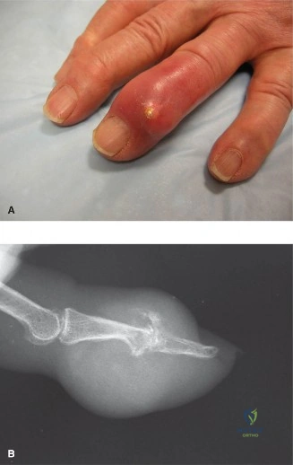
An 82-year-old female presents with a painful, erythematous, and swollen index finger. She has a history of hypertension and renal impairment. Aspiration of the distal interphalangeal joint yields cloudy, chalky fluid. Under polarized light microscopy, you observe the following findings:
What is your primary diagnosis, and what are the three critical steps in the management of this patient?
Candidate: The diagnosis is acute tophaceous gout. The management involves 1) ruling out septic arthritis with cultures, 2) initiating acute anti-inflammatory treatment, and 3) optimizing long-term urate-lowering therapy (ULT).
A poor candidate forgets to address the high likelihood of septic arthritis in the elderly, fails to mention the necessity of checking renal function before prescribing NSAIDs/Colchicine, or suggests immediate surgical excision of the tophus during an active inflammatory phase.
The diagnosis is acute tophaceous gout, confirmed by negatively birefringent needle-shaped crystals. The three-pillar management approach: 1. Rule out Sepsis: Even with crystals, infection can coexist. Perform Gram stain and culture. 2. Acute Inflammatory Control: Start corticosteroids (preferred in elderly/renal patients) or colchicine/NSAIDs (if renal function permits), ensuring systemic coverage. 3. Long-term Systemic Management: Initiate or titrate Urate-Lowering Therapy (e.g., Allopurinol) aiming for an SUA <5 mg/dL, with flare prophylaxis (low-dose colchicine) for the first 3-6 months. Surgical management is reserved for mechanical complications (nerve compression/impending skin rupture) once the acute flare has subsided.
The patient is now quiescent but returns with a large, unsightly tophus at the base of her thumb that is causing triggering of the FPL tendon and median nerve paresthesias. She demands surgery. How do you assess her for surgical suitability?
Candidate: I would assess her current urate levels to ensure she is being treated medically. If her SUA is controlled, I would evaluate the neurological deficit and the risk of skin breakdown. Surgery is indicated because of the functional impairment and nerve compression.
Failing to mention the "Contraindications" list. A failing candidate might operate while the patient has an elevated SUA, which leads to high recurrence and poor wound healing, or ignores the high risk of post-operative gout flares.
Assessment focuses on three areas: 1. Medical Optimization: Verify SUA <6 mg/dL (or <5 mg/dL for tophaceous). If not controlled, surgery is deferred as it significantly increases post-op flare risk and wound complications. 2. Functional/Complication Assessment: Document objective median nerve deficit and FPL excursion. Ensure skin is stable; if ulcerated, surgery is more urgent. 3. Surgical Planning: Ensure a perioperative flare prophylaxis protocol is in place. Discuss the reality that surgery is a "debulking" procedure, not a cure for the systemic metabolic disease. Prioritize longitudinal incisions to preserve vascularity in fragile, aged skin.
