Wassel Type IV Thumb Duplication: Comprehensive Surgical Anatomy, Biomechanics, and Management
Key Takeaway
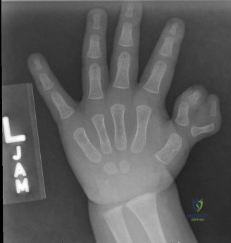
Wassel Type IV thumb duplication involves duplication of the proximal phalanx, with two distinct proximal phalanges articulating with a single metacarpal head. This second most common type presents unique surgical challenges due to its impact on the metacarpophalangeal (MP) joint, requiring detailed understanding of skeletal, ligamentous, tendinous, and neurovascular anomalies for effective reconstruction.
A 9-month-old child presents with congenital thumb duplication. Clinical examination shows a duplicated digit at the metacarpophalangeal (MP) level. Radiographs confirm a shared metacarpal head with two proximal phalanges, each articulating with its own distal phalanx. How would you classify this, and what are the critical systemic associations you must rule out during your preoperative workup?
Candidate: This is a Wassel Type IV thumb duplication. Before surgery, I would perform a systemic evaluation, including a focused history and clinical examination for syndromic associations, specifically looking for Fanconi anemia, VACTERL association, Holt-Oram syndrome, and Diamond-Blackfan anemia. I would likely arrange a pediatric consultation, echocardiogram, and possibly blood work.
Candidates often simply list the syndromes without explaining the rationale. A poor answer ignores the "why"—failing to mention that while most cases are sporadic and isolated, associated congenital anomalies can be life-limiting. They also often fail to mention the need for a formal pediatric review.
Start with the classification: "This is a Wassel Type IV duplication." Then pivot to the systemic check: "While the majority of radial polydactyly is isolated, I must screen for associated syndromes. I would look for physical markers of Fanconi Anemia (e.g., skin pigmentation, short stature), Holt-Oram (cardiac), and VACTERL. I would request a formal pediatric assessment, a baseline echocardiogram to rule out cardiac anomalies (especially relevant in Holt-Oram), and a CBC to screen for marrow failure (Fanconi’s). Any syndromic suspicion requires a multidisciplinary approach before planning elective hand reconstruction."
You have decided to proceed with surgical reconstruction. Describe your decision-making process for choosing which of the two thumb components to retain in a Wassel Type IV case.
Candidate: I would retain the larger, more central, and better-formed thumb. Usually, this is the ulnar thumb. I also look for which thumb has better intrinsic muscle attachments, particularly the Abductor Pollicis Brevis (APB) and the Flexor Pollicis Brevis (FPB), as these are essential for opposition and pinch stability.
Failing to mention the "intrinsic muscle rebalancing." Candidates who focus only on bone/size often forget that the thumb is useless without the motor power (opposition) provided by the intrinsic muscles, which frequently need to be transferred to the retained digit.
Use a structured approach: "I base my decision on three pillars: 1. Skeletal development: Retaining the component with better articular congruity and alignment. 2. Soft tissue: Assessing the neurovascular bundle quality—the retained digit must have a robust supply. 3. Functional balance: The ulnar digit is usually prioritized because it typically possesses superior intrinsic muscle attachments (APB, FPB, Adductor). My surgical plan involves not just excising the radial component, but carefully harvesting the periosteal sleeve and intrinsic insertions from the resected digit to augment the retained thumb’s MP joint stability."
During the procedure, you note that the MP joint is highly unstable after resecting the redundant radial component. What techniques will you employ to ensure a stable, functional joint?
Candidate: I would reconstruct the collateral ligaments, potentially by salvaging the radial collateral ligament from the resected digit and advancing it to the retained thumb. I would also perform a capsulorrhaphy, rebalance the intrinsic muscles, and stabilize the joint with a K-wire for 4-6 weeks.
Missing the mention of the growth plate. A common error is aggressive pinning or dissection that violates the metacarpal epiphysis, risking growth arrest or angulation.
Structure the answer by anatomy: "Stabilization is a three-step process: 1. Ligamentous: I will reconstruct the collateral ligament by harvesting a periosteal flap or ligamentous remnant from the resected digit. 2. Intrinsic: I will perform a formal transfer/reinsertion of the APB/FPB complex to the base of the retained proximal phalanx to provide active dynamic stability. 3. Skeletal: I will use a smooth K-wire to hold the joint in a neutral, anatomic position, ensuring the wire does not cross the physis if avoidable, and is left in situ for 4-6 weeks. I would also consider re-contouring the bifid metacarpal head to a single, smooth articular surface to prevent long-term tracking issues."
