Diagnose Posterior Labrum Tear: Your Interactive Ortho Case
Key Takeaway
Looking for accurate information on Diagnose Posterior Labrum Tear: Your Interactive Ortho Case? A **posterior labrum tear** involves damage to the cartilage rim at the back of the shoulder socket, frequently occurring after a posterior shoulder dislocation. This injury typically happens when the shoulder is flexed, adducted, and internally rotated under trauma. It can cause persistent pain, a sensation of instability, and may include a reverse Bankart lesion, where a bone fragment avulses from the posterior glenoid.
A 22-year-old offensive lineman presents with chronic deep posterior shoulder pain and a sensation of instability. He describes a mechanism of injury involving an axial load applied to his arm while in flexion, adduction, and internal rotation. He endorses a 'dead arm' sensation during bench pressing. Based on the clinical presentation and the provided image, what is your primary differential diagnosis and which provocative tests would you rely upon?
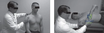
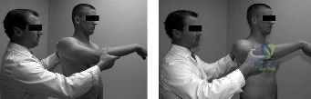
Candidate: Given the history of axial load in the flexed, adducted, and internally rotated position, I am most concerned about a posterior labral tear. The physical exam should include the Jerk test and the Kim test to assess for posterior labral pathology and stability.
Candidates often fail to describe the specific mechanism or confuse posterior instability with anterior instability (e.g., performing an apprehension test instead). They may also forget to mention the need to assess for concomitant conditions like SLAP lesions or scapular dyskinesia.
The patient's clinical presentation—an offensive lineman with 'dead arm' and pain with axial loading in the 'clunk' position—is classic for a posterior labral tear. I would perform the Jerk test (axial load with horizontal adduction) and the Kim test (axial load in 90 degrees elevation with diagonal loading), which have high sensitivity and specificity. I would also perform a Load and Shift test to quantify posterior translation, noting that normal translation is Grade 1, while this patient likely has Grade 2 or greater.
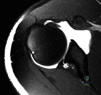
You have ordered an MRA which confirms a posterior labral tear. Looking at the axial MRI slice, how would you classify this pathology, and what specific anatomical variant are you looking for regarding the labrocapsular junction?
Candidate: I would classify this using the Kim Classification, likely a Type II lesion representing a complete detachment. I am specifically evaluating for a POLPSA lesion—a posterior labrocapsular periosteal sleeve avulsion—where the periosteum is stripped but remains intact, creating a redundant capsular recess.
Failing to distinguish between a simple reverse Bankart lesion and a POLPSA lesion. Candidates may also miss the importance of checking glenoid version on the same scan to rule out bony dysplasia.
I would categorize this using the Kim Classification (Types I-IV). Based on the MRI, I am assessing for a POLPSA (Posterior Labrocapsular Periosteal Sleeve Avulsion), where the capsule is stripped off the glenoid neck with the labrum while the periosteum remains attached, creating a "pouch." Differentiating this from a standard reverse Bankart is vital because the surgical approach requires mobilizing that periosteal sleeve to achieve anatomical reduction and adequate tensioning.
The patient has failed 3 months of conservative therapy and you are moving to surgery. Describe your arthroscopic setup, portal selection, and the critical steps for ensuring a robust repair.
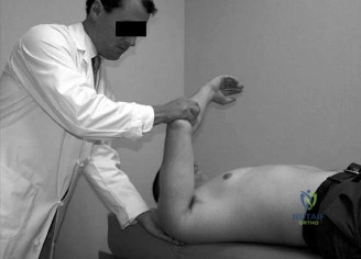
Candidate: I would use the lateral decubitus position for better visualization. I use a standard posterior viewing portal, an anterior working portal, and a dedicated 7 o'clock accessory posterolateral portal. Essential steps include liberating the labrum down to the 6 o'clock position and ensuring the drill guide is parallel to the joint surface to avoid cartilage damage.
Attempting posterior repairs solely through the standard posterior portal. This results in "anchor skiving," where the anchor penetrates the articular surface rather than the bone, and poor vertical orientation of the repair.
Lateral decubitus position is mandatory to facilitate capsular shifting. I employ an accessory 7 o'clock posterolateral portal, which is critical for achieving a perpendicular angle of approach to the glenoid rim. Intraoperatively, I perform extensive mobilization of the labrocapsular complex off the glenoid neck down to the 6 o'clock position. I use all-suture anchors for fixation, taking care to incorporate the posterior band of the inferior glenohumeral ligament (IGHL) for effective capsular plication. Avoiding anchor skiving by ensuring the drill guide is precisely aligned to the articular margin is the highest priority.
Postoperatively, what is the greatest risk in terms of rehabilitation and functional outcome, and how do you monitor for it?
Candidate: The biggest risk is over-tightening the posterior capsule, which leads to a Glenohumeral Internal Rotation Deficit (GIRD). I monitor this by tracking internal rotation ROM during the rehabilitation phases.
Failing to mention the balance between stability and mobility. Suggesting overly aggressive early range of motion, which puts the repair at risk of stretch-out or failure.
The primary concern is the development of a fixed posterior capsular contracture, resulting in a loss of internal rotation (GIRD). This is detrimental for an offensive lineman who requires specific arm positioning for blocking. To mitigate this, I use a phased protocol: absolute immobilization for the first 4 weeks, followed by gradual restoration of motion. I monitor for this by comparing side-to-side internal rotation in the scapular plane and ensuring the patient does not develop anterior impingement symptoms, which are often secondary to excessive posterior tightness.
