Pediatric Glenohumeral Instability: Diagnosis, Anatomy & Management
Key Takeaway
Pediatric glenohumeral instability is symptomatic shoulder laxity or dislocation in skeletally immature patients, particularly adolescents. It's a complex challenge with high recurrence rates due to factors like ligamentous laxity and specific lesions. Diagnosis requires a thorough physical exam, focusing on apprehension signs, and understanding the unique developing anatomy and biomechanics.
A 16-year-old competitive rugby player presents to your clinic after his third episode of anterior shoulder dislocation. His initial injury occurred 18 months ago. He is currently asymptomatic but desires to return to full-contact sport. You perform a physical examination of his shoulder.
Describe the maneuver shown, the findings you would expect to elicit, and the specific clinical question this test is designed to answer.
Candidate: This is the Apprehension test. I would place the patient's arm in 90 degrees of abduction and then externally rotate it. If the patient feels like the shoulder is going to "pop out" or feels apprehensive, the test is positive, which confirms anterior shoulder instability.
Candidates often fail to mention the Relocation Test (Jobe’s). Stopping at the apprehension test ignores the necessary follow-up to differentiate between true anterior instability and generalized guarding or subacromial impingement. Failing to mention the bilateral comparison is also a common omission.
This is the Apprehension test, which assesses for anterior glenohumeral instability. I place the arm in abduction and external rotation; a positive test is marked by patient apprehension or a feeling of impending dislocation. Critically, I would immediately follow this with the Relocation Test: applying a posterior force to the humeral head. If the apprehension disappears, it confirms that the instability is due to anterior capsulolabral incompetence rather than primary subacromial impingement or pain-related guarding. I would also perform this bilaterally to assess for baseline ligamentous laxity.
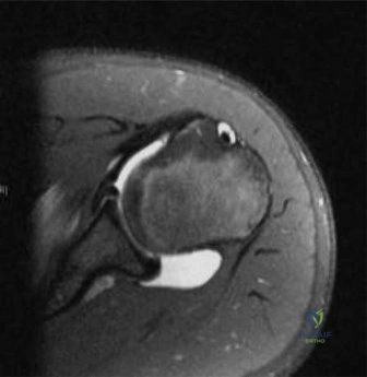
Given the patient's history of three dislocations and his desire to return to contact sports, discuss your threshold for surgical intervention in this adolescent patient versus an adult, and define the "Red Flags" you would look for on imaging that would alter your surgical plan.
Candidate: In an adolescent, the recurrence rate is very high, often cited as over 70%. Therefore, I would have a much lower threshold for surgery than in an adult. I would order an MRI to check for a Bankart lesion and a CT scan if I suspect bone loss.
Failing to quantify bone loss. Stating "I would check for bone loss" without specifying the clinical significance (i.e., the 15-20% threshold) shows a lack of depth. Also, missing the mention of the "engaging" Hill-Sachs lesion is a major gap.
Adolescents face a >70% recurrence rate due to higher activity demands and ligamentous laxity, making surgical intervention for recurrent instability the standard. Regarding imaging, I am looking for specific "Red Flags" that preclude a simple soft-tissue arthroscopic Bankart repair: 1. Glenoid Bone Loss: >15-20% bone loss usually necessitates bony augmentation like a Latarjet procedure. 2. Engaging Hill-Sachs Lesion: A humeral head defect that engages the glenoid during abduction/external rotation, which would require an arthroscopic remplissage or bony augmentation. 3. Significant Capsular Laxity: Identifying this helps determine if a capsular shift or plication is required in addition to labral repair.
