Multidirectional Instability (MDI) of the Shoulder: Anatomy, Biomechanics & Management
Key Takeaway
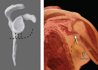
Multidirectional Instability (MDI) of the shoulder is symptomatic laxity in two or more directions (anterior, posterior, inferior) of the glenohumeral joint. Unlike traumatic instability, MDI often arises insidiously from factors like generalized ligamentous laxity, repetitive microtrauma, or neuromuscular dyscoordination, leading to global capsular redundancy and excessive humeral head translation.
A 22-year-old competitive swimmer presents with a 12-month history of global shoulder pain and a subjective sensation of "slipping" during her stroke. She has failed a comprehensive 9-month physiotherapy program. Clinical examination reveals a Beighton score of 6/9 and a positive Sulcus sign. How do you approach the clinical assessment and initial diagnosis of this patient?
Candidate: I would suspect Multidirectional Instability (MDI). I would take a thorough history to rule out trauma, then perform an exam looking for generalized laxity using the Beighton score. I would test for instability in multiple planes using the Sulcus sign, and anterior/posterior load-and-shift tests. I’d order plain films to rule out bony pathology and an MRA to check for labral tears or capsular redundancy.
Focusing solely on the "instability" without discussing the interplay of static and dynamic stabilizers. Failing to specifically mention the importance of distinguishing between symptomatic laxity vs. MDI. Omitting the assessment of scapular dyskinesis, which is often a key contributor in the overhead athlete.
I would approach this by confirming the diagnosis of MDI, characterized by symptomatic laxity in ≥2 directions. My assessment would focus on: 1) Patient Factors: Determining if this is constitutional laxity (Beighton score) vs. acquired/recreational microtrauma. 2) Clinical Exam: Systematic testing (Sulcus, Load-and-Shift) to quantify translation. 3) Dynamic Assessment: Evaluating the rotator cuff and scapular stabilizers, as neuromuscular dyscoordination is a common contributor. 4) Imaging: Ruling out "pseudolaxity" or labral pathology via MRA. I would ensure she has had an adequate trial of neuromuscular-focused physiotherapy before considering her a candidate for surgical stabilization.
Following a failed trial of conservative management, you proceed to arthroscopic intervention. You are currently looking at the glenohumeral joint. Describe your findings and the surgical objective demonstrated in this image.
Candidate: The image shows an arthroscopic view where a suture anchor has been placed on the glenoid neck. The surgeon is capturing redundant capsular tissue to perform a plication. The objective is to reduce the volume of the joint capsule and tighten the inferior glenohumeral ligament (IGHL) complex to improve stability.
Describing this as a "Bankart repair." MDI is a capsular redundancy issue, not a labral detachment issue. Failing to emphasize that thermal capsulorrhaphy is obsolete due to the risk of chondrolysis.
This is an arthroscopic capsular plication or "shift." The surgical goal is to address the global capsular redundancy by folding and securing the attenuated capsule back to the glenoid rim. By doing this inferiorly (addressing the axillary pouch) and potentially anteriorly or posteriorly, we restore the tension of the IGHL complex. Unlike a Bankart repair, we are managing the volume of the capsule itself, not reattaching a labral avulsion.
Your patient is now 3 months post-operative. She complains of persistent stiffness and limited external rotation. How would you manage this patient?
Candidate: I would first reassure the patient, as some stiffness is expected post-capsular plication. I would review her physiotherapy program to ensure we aren't being overly aggressive, but maintain gentle range-of-motion exercises. If it doesn't improve, I might consider an injection or, as a last resort, manipulation under anesthesia.
Suggesting immediate surgical release or manipulation too early. Failing to mention the importance of ruling out other causes of pain or "guarding" that might mimic true mechanical stiffness.
Management must be staged. First, clinical correlation: Is this true capsular contracture or simply guarding due to pain? I would continue supervised physiotherapy with a focus on non-aggressive stretching and modalities. If stiffness persists beyond 6 months and significantly impairs function, I would consider a manipulation under anesthesia (MUA), while remaining cognizant of the risk of disrupting the recent plication. Arthroscopic capsular release is a very rare last resort and carries a high risk of reversing the stability gained by the original procedure.
