Hypoplastic Thumb: Blauth Classification, Surgical Anatomy & Reconstructive Principles
Key Takeaway
Hypoplastic thumb is a congenital anomaly impacting first digit function. It's classified by the Blauth system (Types I-V) based on severity, from mild shortening to complete absence. Surgical goals include deepening the first web space, stabilizing the CMC joint, restoring opposition, and improving pinch strength to maximize hand dexterity and function.
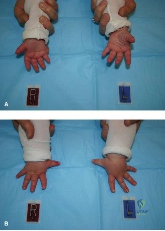
You are presented with an infant with a thumb that appears short and has a restricted first web space. The parents are concerned about the child's ability to grasp objects. You suspect a hypoplastic thumb. How would you classify this and what is the significance of the "Blauth classification" in your management plan?
Candidate: I would use the Blauth classification to grade the severity. Type I is mild, II involves web space narrowing and intrinsic deficiency, III is more severe with CMC instability, and IV/V are floating or absent thumbs. The classification helps decide between reconstruction (Type I-III) or pollicization (Type IV-V, sometimes severe III).
Candidates often confuse Type IIIA and IIIB. They frequently fail to mention the systemic associations (VACTERL/TAR) that must be investigated as part of the initial workup, focusing only on the surgical procedure. They also often forget to mention the functional status of the intrinsic muscles which is critical for decision making.
I would use the Blauth classification as it directly dictates the reconstructive strategy. Type I-II typically require web space release and potential opposition transfer. Type IIIA requires CMC stabilization (capsulodesis/ligament reconstruction) and tendon transfer. Type IIIB (floating) through Type V (aplasia) are managed with pollicization. Crucially, before surgery, I must screen for VACTERL/TAR associations (echocardiogram, renal ultrasound, vertebral assessment), as these systemic anomalies dictate surgical timing and patient safety.
You have decided to proceed with reconstruction for a Blauth Type IIIA thumb. You plan to perform an FDS tendon transfer for opposition. Describe the key technical steps and the importance of tensioning the transfer.
Candidate: I would harvest the FDS from the ring finger, bring it through the palm to the thumb, and secure it to the APB insertion. It is important to tension it correctly so the thumb is in opposition.
Failing to define the required thumb position for tensioning. Simply saying "opposition" is insufficient; the examiner wants to hear the triad of abduction, flexion, and pronation. Missing the need to protect the A2 pulley of the donor finger is a major technical oversight.
The donor is the Ring FDS, harvested at the level of the A2 pulley while maintaining the pulley system to prevent bowstringing. The tendon is routed subcutaneously to the radial-volar aspect of the thumb. The critical step is tensioning: the thumb must be fixed in full abduction, full flexion, and full pronation. This mimics the normal trajectory of the Opponens Pollicis, effectively restoring the mechanical requirements of thumb opposition.
Following your reconstruction of the first web space, the patient returns at 6 months with signs of recurrent adduction contracture. What is your management strategy?
Candidate: I would start them on intense occupational therapy and night splinting. If it doesn't improve, I would perform a revision Z-plasty or consider releasing the adductor muscle.
Assuming the recurrence is purely skin-related. Failing to assess the deep structures (Adductor Pollicis muscle) or the stability of the CMC joint. A passive vs. active contracture must be differentiated.
First, I differentiate between passive skin contracture and active muscular contracture. If passive, I escalate physical therapy with aggressive serial splinting. If the contracture is rigid or fails therapy, surgical revision is needed. This involves a comprehensive release: radical release of the adductor pollicis origin from the 3rd metacarpal, potential capsulotomy of the CMC joint, and if skin remains the limiting factor, a more extensive flap procedure (e.g., dorsal rotation flap or full-thickness skin graft) to ensure adequate soft tissue coverage.
