Elbow Osteoarthritis: Comprehensive Guide to Epidemiology, Anatomy & Clinical Insights
Key Takeaway
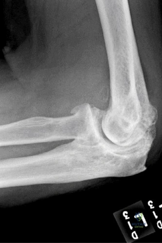
Elbow osteoarthritis is a degenerative joint disease characterized by cartilage loss, pain, and stiffness. While primary OA is less common, most cases are secondary, often stemming from post-traumatic injuries (fractures, dislocations), inflammatory conditions, or repetitive stress. Key features include progressive pain, limited motion, mechanical symptoms, and specific radiographic findings, demanding detailed anatomical understanding for effective management.
A 55-year-old manual laborer presents with progressive elbow pain and stiffness. He describes "locking" and "catching" episodes. On examination, he has a flexion-extension arc of 40°-110°. Radiographs are performed.
How would you classify this clinical presentation, and what are your immediate management considerations?
Candidate: The patient has primary elbow osteoarthritis, likely secondary to his heavy manual labor. The mechanical symptoms and stiffness are characteristic of osteophyte impingement. I would start with non-operative management, including NSAIDs, activity modification, and physical therapy, before considering surgical debridement if he fails to improve.
Failing to mention the ulnar nerve. In a patient with significant osteophytosis and structural deformity, the ulnar nerve is at high risk of secondary entrapment. A candidate who ignores the neurological assessment of the ulnar nerve will score poorly.
This is symptomatic elbow osteoarthritis with secondary mechanical block. My assessment would be: 1) Evaluate for ulnar neuropathy clinically and electromyographically; 2) Confirm failure of 3-6 months of conservative management; 3) If surgical, propose arthroscopic or open debridement (Outerbridge-Kashiwagi) to address osteophyte impingement, noting that the primary goal is restoration of the functional arc of motion (30°-130°) and clearance of loose bodies.
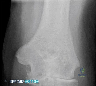
You have decided to proceed with a Total Elbow Arthroplasty (TEA) for a patient with end-stage rheumatoid arthritis. The patient has significant bone loss.
What are the critical intraoperative "no-go" zones or structures at risk during this approach, and how do you protect them?
Candidate: The most important structure is the ulnar nerve, which must be identified and protected. The radial nerve is also at risk, especially if performing a lateral approach. I would use a posterior approach and ensure the ulnar nerve is transposed if necessary.
Neglecting the triceps mechanism. In TEA, the triceps is the power source for extension. If the triceps detachment/repair is handled poorly, the patient will have a significant extension lag post-operatively, which is a major functional failure.
The primary at-risk structures are the ulnar nerve (identify early, protect in a vessel loop, and perform anterior transposition if tension or displacement is likely) and the triceps mechanism. I prefer a triceps-sparing approach (Bryan-Morrey or similar) to preserve the extensor mechanism. If a triceps-splitting or reflecting approach is used, a meticulous, bone-to-bone repair is required to avoid an extension lag. I also monitor the radial nerve if approaching laterally.
The patient has now undergone a successful TEA.
What are the absolute, non-negotiable post-operative instructions you provide this patient, and why?
Candidate: I would tell them not to lift anything too heavy. Generally, they should avoid heavy work and impact activities to ensure the implant stays fixed to the bone and doesn't loosen.
Vague instructions like "don't lift too heavy" are insufficient for an FRCS level. Candidates must provide specific numerical limits and the underlying biomechanical rationale (implant-bone interface failure).
The patient requires strict, lifelong weight-bearing restrictions: no lifting >10-15 lbs (approx. 5 kg) and no repetitive pushing/pulling or impact activities. The rationale is that TEA has a high rate of aseptic loosening due to the significant torque forces at the implant-bone interface. Protecting the link and the cement-bone bond is essential to prevent periprosthetic fracture and early implant failure.
