Bennett's Fracture-Dislocation: Clinical Presentation, Diagnostics & Management
Key Takeaway
Bennett's fracture-dislocation is an intra-articular fracture of the first metacarpal base, with a small volar-ulnar fragment and dorsal-radial-proximal displacement of the main shaft. Diagnosis involves clinical findings (pain, instability) and confirmed by specialized X-rays (Robert's view) and CT scans for precise articular evaluation and surgical planning.
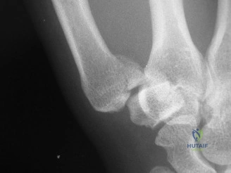
A 32-year-old male presents with acute pain at the base of his thumb after punching a wall. Examination reveals tenderness at the CMC joint and a thumb-in-palm deformity. You are presented with the following radiograph. Describe your findings and the biomechanical forces acting on this fracture.
Candidate: This is a Bennett fracture-dislocation. There is a small volar-ulnar fragment attached to the trapezium, and the main metacarpal shaft is displaced proximally, dorsally, and radially. This is caused by the pull of the Abductor Pollicis Longus (APL) on the shaft, while the fragment stays in place because of the Anterior Oblique Ligament.
Failing to mention the "Anterior Oblique Ligament" (or "beak" ligament) by name, or ignoring the dynamic contribution of the Adductor Pollicis, which creates the rotational/supination torque that complicates the reduction.
Identify the injury as an intra-articular fracture-subluxation. State that the volar-ulnar (Bennett) fragment is tethered to the trapezium by the intact anterior oblique ligament. Explain the displacement of the metacarpal shaft as a result of the APL (proximal/radial/dorsal pull) and the Adductor Pollicis (ulnar/supination torque). Finally, define the goal of treatment as anatomical restoration of the articular surface to prevent post-traumatic CMC osteoarthritis.
You decide to take this patient to theatre. What specific reduction maneuver do you perform to achieve a stable position prior to fixation?
Candidate: I would apply longitudinal traction to the thumb, then place it in palmar abduction and pronate the metacarpal, while pushing the base of the metacarpal dorsally and ulnarly to reduce it against the Bennett fragment.
Forgetting to specify the pronation component of the reduction, which is vital because the Adductor Pollicis induces a supination torque on the metacarpal shaft.
Structure the answer clearly: 1. Longitudinal traction to overcome APL shortening. 2. Palmar abduction to tighten the capsuloligamentous structures. 3. Pronation of the metacarpal to correct the rotational malalignment. 4. Direct pressure (or 'joystick' manipulation) to translate the metacarpal base ulnarly into the saddle joint.
During your surgical approach, you are concerned about injury to the sensory nerves. Specifically, which nerves are at risk, and how do you protect them?
Candidate: The superficial branch of the radial nerve is at risk dorsally, and the lateral antebrachial cutaneous nerve is at risk volarly. I protect them by performing careful, blunt dissection in the subcutaneous layer and identifying the internervous plane.
Simply saying "I am careful" without naming the specific nerves or the layers where they are located. Failing to identify the internervous plane between the radial-nerve innervated APL/EPB and the median-nerve innervated thenar muscles.
Identify the superficial radial nerve (dorsal) and lateral antebrachial cutaneous nerve (volar). Emphasize that the Wagner approach requires meticulous dissection under loupe magnification, identifying these structures early, and staying strictly within the internervous plane to avoid inadvertent retraction or traction injuries.
