Soft Tissue Mallet Finger: Comprehensive Review of Anatomy, Classification, & Management
Key Takeaway
Soft tissue mallet finger, a Doyle Type I injury, involves terminal extensor tendon disruption at the DIP joint, causing a flexion deformity. It's a common hand injury, typically managed non-operatively with continuous DIP joint splinting in extension for 6-8 weeks to allow tendon healing and prevent secondary deformities like swan neck.
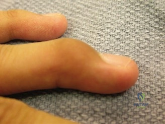
A 35-year-old amateur rugby player presents with a "jammed" middle finger sustained during a match 4 days ago. On examination, he has a 45-degree extensor lag at the DIP joint. Passive extension is full. Radiographs are provided below. What is your diagnosis, classification, and management strategy?
Candidate: "This is a closed, acute soft tissue mallet finger. Based on the Doyle classification, it is a Type I injury. Given that it is acute and passively correctable, I would manage this non-operatively with continuous splinting of the DIP joint in neutral or slight hyperextension for 6 to 8 weeks, leaving the PIP joint free."
Candidates often fail to explicitly mention the "continuity" of the splinting—a common reason for failure is the patient removing it to wash. Also, failing to mention the assessment of the PIP joint or not verifying that the injury is truly a 'soft tissue' type (ruling out bony avulsion on the radiograph) demonstrates a lack of diagnostic rigor.
The candidate should state: "Clinical diagnosis is a Doyle Type I soft tissue mallet finger. My management is non-operative: 24/7 continuous DIP extension splinting for 6–8 weeks. I would emphasize to the patient that any flexion of the DIP joint resets the clock on healing. I will ensure the PIP joint is free to move to prevent stiffness. I would follow up at 2 weeks to ensure skin integrity and splint compliance, and finally, warn the patient about the potential for a mild permanent extensor lag, which is a known outcome even with perfect compliance."
The patient returns at 10 weeks. He admits to removing the splint to shower every day. Clinical assessment now shows a 25-degree fixed extensor lag at the DIP joint and an early hyperextension deformity of the PIP joint. How do you explain the secondary deformity, and what is your plan?
Candidate: "The patient has developed a symptomatic secondary swan-neck deformity. This is due to the loss of the terminal extensor tendon, which leads to proximal migration of the lateral bands. These bands shift dorsally to the axis of the PIP joint, causing hyperextension. I would recommend a trial of PIP flexion splinting and continued nighttime DIP extension, but if this fails to improve the lag or the swan-neck, I would discuss surgical options, such as DIP fusion or reconstructive tenodesis."
Suggesting immediate surgery for a 25-degree lag. A borderline candidate ignores the patient's functional deficit and rushes to suggest aggressive surgery for a deformity that might still respond to therapy or may be acceptable to the patient.
A high-scoring answer addresses the biomechanics: "The swan-neck deformity is a result of the terminal extensor deficit causing a lateral band shift. I would categorize the deformity as 'flexible' or 'fixed' on exam. If flexible, I will prioritize physical therapy for PIP flexion and nighttime DIP splinting. I would avoid invasive surgery unless the patient has a significant functional deficit or the deformity becomes fixed and painful, at which point I would consider DIP arthrodesis or soft tissue reconstruction."
