Jersey Finger: Tendon Avulsion at the Base of the Distal Phalanx
Key Takeaway
Learn more about Jersey Finger: Tendon Avulsion at the Base of the Distal Phalanx and how to manage it. A "jersey finger" is an avulsion injury where the flexor profundus tendon detaches from its insertion at the base of the distal phalanx. This injury typically prevents the patient from fully flexing the distal interphalangeal (DIP) joint and often presents with pain, swelling, and bruising, especially after a forceful hyperextension injury. Early surgical repair is usually recommended.
A 24-year-old rugby player presents with acute pain and swelling at the base of his ring finger following a tackle. He is unable to flex the DIP joint. You suspect an FDP avulsion. Describe your initial clinical assessment and what radiographic findings you are looking for.
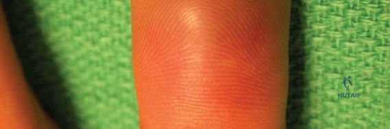
Candidate: I would examine the patient by isolating the FDP. I hold the MCP and PIP joints in extension and ask the patient to flex the DIP joint; inability to do so confirms the diagnosis. I would then order AP, lateral, and oblique radiographs of the finger to look for a bony avulsion fragment, which would help me classify the injury according to the Leddy and Packer system.
Failure to specifically mention the stabilization of the PIP and MCP joints. Without neutralizing the FDS, a patient might demonstrate residual flexion, leading to a missed diagnosis. Also, failing to mention palpating for the retracted stump in the palm is a common oversight.
The candidate systematically isolates the FDP by holding the MCP and PIP in full extension to negate FDS function. They note the need for clinical palpation of the palm to check for a retracted tendon nodule (indicating Type I). Radiographically, they prioritize identifying the size and location of bony fragments to differentiate between Type III (large fragment) and Type IV (double fragment) injuries, noting that lateral views are essential to see volar displacement relative to the A4 pulley.
You have diagnosed a Type I Jersey finger. Explain the significance of the vincula system in this specific classification and why it dictates the urgency of your surgical management.
Candidate: In a Type I injury, the FDP has retracted into the palm. This means both the vinculum longum and the vinculum brevis are ruptured, which cuts off the primary blood supply to the tendon. Because the tendon is essentially devascularized, it is a surgical emergency; we should operate within 7 to 10 days to prevent tendon necrosis and irreversible shortening.
Focusing only on the clinical timing without explaining the *pathophysiology* (vascular supply via vincula). Failing to link the "retraction into the palm" to the rupture of both vincula is a missed opportunity to show deeper understanding.
The candidate explicitly links the anatomy (the dual-source blood supply: synovial diffusion and the vincula system) to the classification. They correctly state that Type I represents a "watershed" injury where the vincula are disrupted, leading to ischemia. They emphasize that the 7-10 day window is not just for preventing scarring, but for salvaging a devascularized structure before irreversible necrosis occurs.
You are in the OR for a Type II FDP avulsion. You have made a Brunner incision and identified the tendon retracted to the level of the PIP joint. What are your priorities when retrieving and reattaching the tendon, and how do you avoid the "Quadriga effect"?
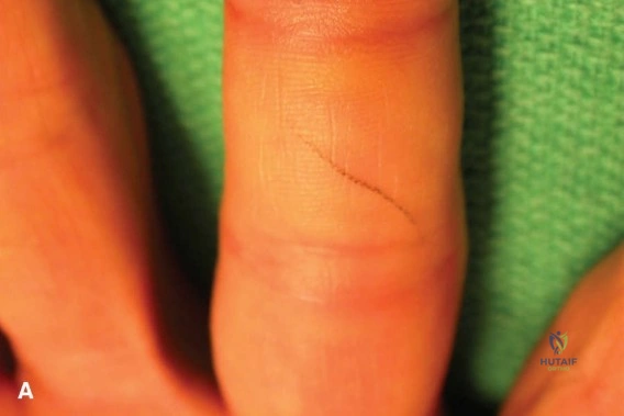
Candidate: My priority is to preserve the A2 and A4 pulleys to prevent bowstringing. I will use a tendon passer to retrieve the FDP through the sheath. To avoid the Quadriga effect, I must ensure I do not overtighten the tendon; I should match the tension to the contralateral uninjured digit, generally avoiding advancing the tendon more than 1cm from its original resting length.
Forgetting to mention the specific pulleys (A2 and A4). Also, failing to explain how the Quadriga effect happens (the FDP tendons share a common muscle belly in the forearm, so shortening one limits the total excursion of the others).
The candidate emphasizes meticulous handling—using only the tip of the tendon to avoid epitenon damage. They identify A2 and A4 as biomechanically critical. They define the Quadriga effect clearly: because the ring FDP shares a muscle belly with the middle and small fingers, over-tensioning the repair creates a relative shortening that prevents full composite flexion of the adjacent fingers. They suggest intraoperative assessment by comparing resting posture and excursion against adjacent digits.
Look at this radiograph of a Type III Jersey finger. Why is fixation of the bony fragment mandatory, and how does your management approach differ from a soft-tissue avulsion?
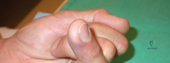
Candidate: Fixation is mandatory to restore DIP joint congruency and tendon function. Unlike a soft-tissue avulsion, where I would use a suture anchor into the bone, here I must anatomically reduce the bone fragment itself. I would use mini-screws or parallel K-wires to stabilize the fragment, ensuring the FDP remains attached to the fragment.
Treating the bony fragment like a standard soft-tissue repair. If you simply anchor the tendon and ignore a large articular fragment, you leave the patient with a step-off in the DIP joint and an inevitable post-traumatic arthritis.
The candidate prioritizes joint surface reconstruction. They explain that the fragment is often large enough to cause joint incongruity and instability. They correctly distinguish that while suture anchors work for Type I/II, Type III requires rigid internal fixation (screws or tension band) to restore the anatomy. They also mention that the FDP's integrity to the bone fragment must be checked; if it has pulled off the fragment (Type IV), that requires a hybrid repair of both bone and tendon.
