Acute Median Nerve and Flexor Tendon Injury: A Distal Forearm Laceration Case Study
Key Takeaway
An acute distal forearm laceration often presents with profound sensory deficits in the median nerve distribution (thumb, index, middle, radial ring fingers) and specific motor deficits like inability to flex the thumb IP, index/middle DIP joints, or abduct the thumb. Diagnosis involves detailed clinical neurological and musculoskeletal assessment, supported by imaging to rule out fractures or foreign bodies.
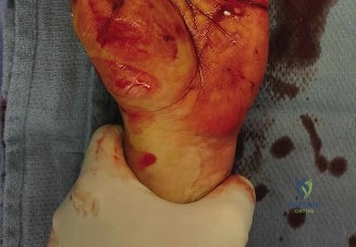
You are in the trauma bay. A 32-year-old male presents with a 5cm oblique volar distal forearm laceration following a glass injury. He has an obvious "abnormal cascade" of the digits. Given the clinical presentation shown, describe your immediate assessment and explain the significance of the "abnormal cascade" findings.
Candidate: I would start by ensuring hemodynamic stability and examining the neurovascular status. The "abnormal cascade"—where the index and middle fingers are extended compared to the others—indicates a loss of the normal resting flexion of the FDS and FDP tendons. This suggests a transection of those flexor tendons. I would also perform a motor and sensory exam to rule out median nerve injury, given the location in Zone V.
Focusing only on the tendon injury while ignoring the possibility of occult skeletal issues or vascular compromise. Failing to emphasize that the resting hand posture is an objective, non-provocative way to confirm tendon disruption without causing the patient pain.
A systematic approach: 1) Vascular: Assess radial/ulnar pulses and perfusion. 2) Neurological: Document sensation in the median nerve distribution and motor function of the APB/opponens before any local anesthetic or sedation. 3) Musculoskeletal: Interpret the "abnormal cascade" as clinical evidence of proximal retraction of the FDS/FDP tendons. Mention that this finding is pathognomonic for complete tendon rupture, negating the need for aggressive pain-inducing range of motion testing in the initial triage phase.
Radiographs confirm the presence of retained radiopaque foreign bodies. How do you approach the surgical planning for this "spaghetti wrist," and what are the specific pearls for the intraoperative repair of these structures in Zone V?
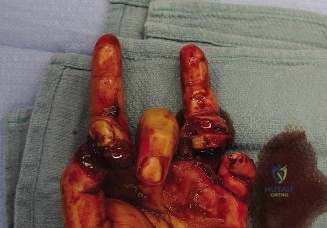
Candidate: I would plan for an urgent, formal surgical exploration under tourniquet control. I would use Brunner incisions to extend the wound, identify all structures systematically, and remove the foreign bodies under C-arm guidance. I would perform core suture tendon repairs with an epitendinous stitch and a tension-free epineurial nerve repair under the microscope.
Failing to mention the sequence of repair (deepest structures first). Not discussing the importance of the epitendinous suture for gliding, or forgetting to mention the need for prophylactic fasciotomy or leaving the antebrachial fascia open in cases of significant swelling.
Structure the answer by: 1) Exposure: Longitudinal zigzag extensions to prevent flexion contractures. 2) Debridement: Fluoroscopic guidance for foreign body removal. 3) Sequence: Repair deepest structures (FDP/FPL) first to avoid working over already repaired tendons. 4) Technique: Use a 4-strand modified Kessler core suture with a 6-0 epitendinous running suture. 5) Nerve: Microscopic epineurial repair using 8-0 monofilament, prioritizing fascicular rotational alignment using vascular landmarks. 6) Post-op: Proactive dorsal blocking splinting and a strict supervised therapy protocol.
