Jersey Finger: Tendon Avulsion at the Base of the Distal Phalanx

Key Takeaway
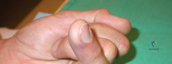
Learn more about Jersey Finger: Tendon Avulsion at the Base of the Distal Phalanx and how to manage it. A "jersey finger" is an avulsion injury where the flexor profundus tendon detaches from its insertion at the base of the distal phalanx. This injury typically prevents the patient from fully flexing the distal interphalangeal (DIP) joint and often presents with pain, swelling, and bruising, especially after a forceful hyperextension injury. Early surgical repair is usually recommended.
Introduction and Epidemiology
Jersey finger refers to an avulsion injury of the flexor digitorum profundus (FDP) tendon from its insertion at the palmar base of the distal phalanx. This injury typically occurs when the FDP tendon is subjected to forceful eccentric contraction while the distal interphalangeal (DIP) joint is rapidly and forcibly extended. The classic mechanism involves a finger, often the ring finger, catching on an opponent's jersey during athletic activities, leading to forced extension of an actively flexed DIP joint while the proximal interphalangeal (PIP) joint and metacarpophalangeal (MCP) joint remain relatively flexed. This dynamic places maximal stress on the FDP insertion.
While commonly associated with American football, Jersey finger can occur in various sports and occupational settings involving forceful gripping and tearing motions. The ring finger is most frequently affected due to its relative laxity and tendency to be more exposed during gripping activities. The injury can occur in isolation or, less commonly, as part of a more complex hand trauma.
Clinical presentation typically includes acute pain at the palmar aspect of the DIP joint, swelling, and ecchymosis. The hallmark sign is the inability of the patient to actively flex the DIP joint in isolation while the MCP and PIP joints are held in extension. Palpation may reveal tenderness at the base of the distal phalanx, and a palpable mass or fullness may be present in the palm if the tendon has retracted significantly. Early diagnosis and intervention are critical for optimal functional outcomes.

Leddy and Packer Classification System
The most widely accepted classification system, introduced by Leddy and Packer in 1977, categorizes FDP avulsions based on the degree of tendon retraction and the presence of a bony avulsion fragment. This classification dictates the urgency and surgical approach:
- Type I: The FDP tendon is completely avulsed from the distal phalanx and retracts into the palm. This occurs because the vinculum longum is torn, compromising the tendon's blood supply and tethering. These are considered surgical emergencies due to the risk of tendon ischemia and permanent shortening. Repair is mandated within 7 to 10 days.
- Type II: The FDP tendon retracts to the level of the PIP joint. The vinculum longum may be partially intact, providing some tethering and maintaining a blood supply. While less urgent than Type I, repair within 2 to 3 weeks is generally recommended to prevent further retraction and contracture.
- Type III: A large bony fragment is avulsed with the FDP tendon, which typically retracts to the level of the A4 pulley or proximal to the A3 pulley, blocked by its size. The fragment is often large enough to cause joint incongruity. This type requires surgical repair to restore joint stability and tendon function.
- Type IV: Described by Smith in 1981, this involves an avulsion of the FDP tendon and a small bony fragment, with the tendon itself having pulled through the bony fragment. This results in two fragments: the small piece attached to the tendon, which retracts, and a larger fragment left attached to the distal phalanx.
- Type V: An epiphyseal avulsion in skeletally immature patients, recently added to the classification, often treated similarly to Type III depending on displacement.
Understanding these types is paramount for guiding management decisions, particularly concerning surgical timing and technique.
Surgical Anatomy and Biomechanics
A thorough understanding of the flexor tendon anatomy and biomechanics is fundamental for successful repair of Jersey finger.
Flexor Digitorum Profundus Tendon Anatomy
The FDP originates from the volar and medial aspects of the ulna and the adjacent interosseous membrane. Its muscle belly lies deep to the flexor digitorum superficialis (FDS) in the forearm. In the wrist, the four FDP tendons pass through the carpal tunnel, deep to the FDS tendons. Within the hand, each FDP tendon enters its respective fibrous flexor sheath at the level of the metacarpal neck.
The fibrous flexor sheath is composed of annular (A1-A5) and cruciate (C1-C3) pulleys. The annular pulleys provide the critical mechanical advantage required for digital flexion by keeping the tendon closely apposed to the phalanges, thereby preventing bowstringing. The A2 pulley (located over the proximal phalanx) and the A4 pulley (located over the middle phalanx) are the most biomechanically critical and must be preserved or reconstructed during surgical intervention to maintain functional tendon excursion and force transmission.

Vascular Supply and the Vincula System
The blood supply to the flexor tendons within the digital sheath is dual-source: vascular perfusion and synovial diffusion. Vascular perfusion is delivered via the segmental vincula system.
* Vincula Brevia: Located near the insertions of the FDS and FDP.
* Vincula Longa: Located more proximally, tethering the tendons to the dorsal aspect of the sheath.
In a Type I Jersey finger, the violent avulsion ruptures both the vinculum longum and brevis of the FDP, completely devascularizing the distal tendon. The tendon subsequently retracts into the palm, relying solely on limited diffusion. This rapid ischemia leads to tendon necrosis and contraction, explaining why Type I injuries represent a true hand surgery emergency requiring intervention within 7 to 10 days. In contrast, Type II and III injuries maintain variable degrees of intact vincula, preserving vascularity and allowing for a slightly delayed surgical window.
Biomechanical Susceptibility of the Ring Finger
The disproportionate incidence of Jersey finger in the ring digit (comprising approximately 75% of cases) is rooted in its unique biomechanical constraints. The FDP muscle bellies to the middle, ring, and small fingers share a common muscle origin and tend to act as a single functional unit (the quadriga effect). During a gripping motion (such as grabbing a jersey), if the opponent pulls away, the middle and small fingers often slip off, extending fully. The ring finger, however, remains engaged. Because the ring finger FDP is anatomically tethered to the adjacent extending digits, its independent excursion is severely limited. Consequently, the ring finger FDP absorbs the entirety of the eccentric load, exceeding the tensile strength of the tendon-bone interface and resulting in avulsion.
Indications and Contraindications
Acute FDP avulsions are almost universally managed operatively to restore distal interphalangeal joint flexion and grip strength. Non-operative management is highly restricted and generally reserved for specific patient populations or delayed presentations where primary repair is no longer feasible.
| Clinical Scenario | Operative Management | Non-Operative Management |
|---|---|---|
| Acute Presentation (< 3 weeks) | Absolute indication for Types I, II, III, IV, and V. Primary repair or bony fixation. | Contraindicated in active patients. |
| Delayed Presentation (3 - 6 weeks) | Relative indication. Primary repair may be attempted if tendon excursion permits without excessive tension. | Considered if severe contracture is present, or if the patient accepts DIP joint functional deficit. |
| Chronic Presentation (> 6 weeks) | Indicated for symptomatic patients. Options include DIP arthrodesis, two-stage tendon reconstruction, or tenodesis. | Acceptable in low-demand patients or those who have adapted to the isolated DIP flexion deficit. |
| Patient Factors | High-demand athletes, manual laborers, young patients. | Medically unfit for surgery, severe peripheral vascular disease, non-compliant patients. |
| Joint Status | Congruent, mobile DIP joint. | Pre-existing severe osteoarthritis of the DIP joint (favors primary arthrodesis). |
Pre Operative Planning and Patient Positioning
Thorough preoperative planning is essential to determine the level of tendon retraction, the presence of bony fragments, and the necessary surgical equipment.
Clinical Evaluation and Imaging
Physical examination must isolate the FDP. The examiner holds the patient's MCP and PIP joints in strict extension to neutralize the FDS tendon; the patient is then asked to actively flex the DIP joint. Inability to do so confirms FDP discontinuity. Careful palpation along the volar digit and palm can often identify the retracted tendon stump, which presents as a tender nodule.
Standard radiographic evaluation includes true anteroposterior (AP), lateral, and oblique views of the affected digit.

Radiographs are critical for identifying Type III and Type IV injuries. A bony fragment visualized volar to the middle phalanx (A4 pulley) or proximal phalanx (A2 pulley) dictates the surgical approach. In cases where the tendon retraction level is clinically ambiguous and no bony fragment is visible, high-resolution ultrasonography or Magnetic Resonance Imaging (MRI) without contrast can precisely locate the retracted tendon stump, aiding in incision planning.
Patient Positioning and Equipment
- Positioning: The patient is positioned supine with the affected upper extremity extended on a radiolucent hand table.
- Anesthesia: Regional anesthesia (axillary or supraclavicular brachial plexus block) or general anesthesia is preferred. Wide-awake local anesthesia no tourniquet (WALANT) is increasingly utilized, allowing for intraoperative active motion testing to assess repair strength and gap formation.
- Tourniquet: A well-padded pneumatic arm tourniquet is applied (unless WALANT is used) to ensure a bloodless field.
- Magnification: Surgical loupes (minimum 2.5x to 3.5x magnification) are mandatory for identifying neurovascular structures and ensuring meticulous tendon handling.
- Equipment: Mini C-arm fluoroscopy, precise soft tissue retractors (e.g., Ragnell, Senn), tendon passers or pediatric feeding tubes, suture anchors (micro-anchors), buttons for pull-out techniques, and appropriate core suture material (e.g., 3-0 or 4-0 non-absorbable braided suture).
Detailed Surgical Approach and Technique
The surgical objective is to retrieve the retracted FDP tendon, pass it anatomically through the flexor sheath without compromising the critical A2 and A4 pulleys, and secure it robustly to the distal phalanx base.
Surgical Incision and Exposure
The approach utilizes a volar Brunner (zigzag) incision or a mid-lateral incision over the affected digit.

The Brunner incision is meticulously designed so that the apices of the flaps do not cross the flexion creases at a 90-degree angle, preventing postoperative scar contracture. Full-thickness fasciocutaneous flaps are elevated, taking care to identify and protect the paired volar digital arteries and nerves, which lie immediately deep to the flaps. The fibrous flexor sheath is exposed from the A1 pulley to the distal phalanx.
Tendon Retrieval
If the tendon has retracted into the palm (Type I), a separate transverse or Brunner incision is made in the distal palmar crease over the A1 pulley. The FDP stump is identified. It is critical to handle the tendon only by its very distal tip to avoid crushing the epitenon, which can exacerbate adhesion formation.

A flexible tendon passer or a small pediatric feeding tube is introduced retrogradely from the distal sheath opening (distal to the A4 pulley), passed under the A4, C pulleys, and A2 pulley, and retrieved in the palm. A core suture is placed in the FDP stump, passed through the feeding tube, and the tendon is gently delivered back to its insertion site. If the tendon is swollen and cannot pass through the chiasm of Camper or the A4 pulley, a limited venting of the A4 pulley or the cruciate pulleys may be performed, though the A2 must remain strictly intact.
Reduction and Fixation Techniques
Preparation of the insertion site requires debridement of the volar base of the distal phalanx to bleeding cancellous bone to promote tendon-to-bone healing.
1. Suture Anchor Repair (Soft Tissue Avulsion - Types I and II):
Currently, the most common technique involves the use of micro-suture anchors. One or two small anchors (e.g., 1.0mm to 1.5mm) are placed into the volar base of the distal phalanx.

A locking core suture (e.g., modified Kessler or Krackow technique) is placed into the distal end of the FDP tendon. The sutures from the anchor are passed through the tendon, and the tendon is advanced to the bony footprint and tied securely. This technique avoids the complications associated with dorsal buttons.
2. Pull-Out Button Technique (Classic Bunnell Technique):
Historically the gold standard, this involves placing a locking core suture into the FDP, passing both suture limbs through drill holes made from the volar base of the distal phalanx exiting the dorsal nail matrix or distal fingertip. The sutures are then tied over a sterile button resting on the dorsal skin. While providing excellent pull-out strength, this method carries risks of nail bed deformity, dorsal skin necrosis, and infection at the button site.
3. Bony Fragment Fixation (Type III):
If a large bony fragment is present, anatomic reduction is required. The fragment is reduced into its crater at the base of the distal phalanx. Fixation is typically achieved using multiple parallel 0.028-inch or 0.035-inch Kirschner wires, a mini-screw (e.g., 1.3mm or 1.5mm), or a tension-band construct. The integrity of the FDP insertion onto the fragment must be verified; if compromised (Type IV), simultaneous tendon-to-bone repair is necessary.
Closure
Following fixation, the tourniquet is deflated, and hemostasis is achieved. The skin is closed with non-absorbable monofilament sutures (e.g., 5-0 nylon). A sterile dressing is applied, and the extremity is immobilized in a dorsal blocking splint.
Complications and Management
Surgical management of Jersey finger is technically demanding, and complications are relatively common. Meticulous surgical technique and strict adherence to postoperative rehabilitation are imperative to mitigate these risks.

| Complication | Incidence / Risk Factors | Management and Salvage Strategies |
|---|---|---|
| DIP Joint Stiffness / Flexion Contracture | Most common complication. Caused by prolonged immobilization or aggressive scarring. | Aggressive hand therapy. If refractory >6 months, tenolysis or capsulotomy may be indicated. Severe cases may require DIP arthrodesis. |
| Tendon Rupture / Repair Failure | 3% - 9%. Often due to patient non-compliance, premature active loading, or technical failure of the core suture. | Requires immediate re-operation. If acute, revision primary repair. If delayed with retraction, two-stage reconstruction or arthrodesis. |
| Quadriga Effect | Occurs if the FDP is advanced too far distally (over-tensioned) during repair, limiting excursion of adjacent digits. | Prevention is key (do not advance tendon >1cm). If symptomatic, surgical lengthening or tenolysis is required. |
| Nail Bed Deformity | Associated primarily with the dorsal pull-out button technique due to iatrogenic injury to the germinal matrix. | Avoidance of dorsal button when possible. If present, nail bed reconstruction may be attempted, though results vary. |
| Infection / Button Necrosis | < 5%. Risk increased with dorsal pull-out buttons or prolonged surgery. | Oral or IV antibiotics. Prompt removal of the button if necrosis occurs. Debridement if deep infection is suspected. |
| Lumbrical Plus Deformity | Occurs if the FDP graft or repair is too loose, causing paradoxical PIP extension during attempted active flexion. | Lumbrical division (release) to restore normal biomechanics. |
Post Operative Rehabilitation Protocols
Rehabilitation is arguably as critical as the surgical repair itself. The goal is to facilitate tendon gliding to prevent restrictive adhesions while protecting the healing tendon-bone interface from excessive tension that could lead to gap formation or rupture.

Immobilization Phase (Weeks 0-4)
Immediately postoperatively, the hand is placed in a dorsal blocking splint (DBS). The wrist is positioned in 20-30 degrees of flexion, the MCP joints in 70-80 degrees of flexion, and the PIP/DIP joints in neutral (0 degrees). This position maximizes relaxation of the flexor musculotendinous unit.
Depending on the strength of the repair and the surgeon's preference, protocols vary between:
* Modified Kleinert Protocol: Utilizes dynamic rubber band traction to maintain passive flexion, allowing active extension against the bands within the confines of the splint.
* Modified Duran Protocol: Relies on strict passive range of motion exercises performed by the patient or therapist, without dynamic traction.
* Early Active Motion (Place and Hold): Increasingly popular for robust repairs (e.g., 4-strand or 6-strand core sutures + anchors). The patient passively flexes the digits and gently actively holds the fist position.
Intermediate Phase (Weeks 4-8)
At 4 weeks, the dorsal blocking splint is typically discontinued during the day and replaced with a static night splint. Active range of motion exercises are initiated. Tendon gliding exercises (straight fist, hook fist, composite fist) are emphasized to maximize differential glide between the FDS and FDP. Blocking exercises to isolate DIP flexion begin around week 6.
Strengthening and Return to Play (Weeks 8-12+)
Progressive strengthening begins at 8 weeks with putty and grip exercises. Unrestricted activity and return to contact sports are generally not permitted until 10 to 12 weeks postoperatively, once the tendon-to-bone healing is mechanically mature.
Summary of Key Literature and Guidelines
The management of Jersey finger remains heavily guided by foundational anatomical studies and evolving biomechanical research regarding fixation constructs.
- Leddy JP, Packer JW (1977): The seminal paper establishing the classification system for FDP avulsions. Their correlation of the level of tendon retraction with the integrity of the vincula system remains the cornerstone of surgical decision-making and timing.
- Smith JH (1981): Expanded the classification to include Type IV injuries, highlighting the necessity of recognizing simultaneous bony avulsion and tendon pull-through to avoid catastrophic failure of isolated bony fixation.
- Biomechanical Fixation Studies (e.g., Silva et al., McCallister et al.): Modern literature has extensively compared suture anchors to traditional pull-out buttons. Current consensus demonstrates that suture anchors provide equivalent or superior biomechanical pull-out strength while significantly mitigating the risks of dorsal skin necrosis, nail bed deformity, and pin-tract infections associated with button techniques. Suture anchors are now considered the gold standard for Type I and II repairs.
- Rehabilitation Evolution: Recent guidelines favor early active motion (EAM) protocols over traditional passive protocols, provided a multi-strand repair or robust anchor fixation is achieved. EAM has been shown to decrease the incidence of debilitating postoperative stiffness without significantly increasing the risk of tendon rupture.