Advanced Rheumatoid Arthritis of the Elbow: Pathophysiology, Anatomy, and Treatment Strategies
Key Takeaway
Advanced rheumatoid arthritis in the elbow involves progressive joint destruction, pain, and functional impairment, often requiring surgical intervention when medical management fails. Key considerations include detailed understanding of the elbow's complex surgical anatomy, biomechanics, and neurovascular structures, alongside careful evaluation of specific indications for procedures like synovectomy or total elbow arthroplasty.
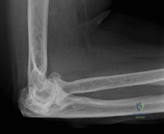
You are presented with a 58-year-old female patient with a 15-year history of seropositive rheumatoid arthritis. She complains of severe, bilateral elbow pain, significant stiffness, and an inability to perform basic perineal care. She has failed a prolonged course of DMARDs and recent biologic therapy. On examination, she has a fixed flexion deformity of 45 degrees, valgus instability, and palpable crepitus. Radiographs are shown below.
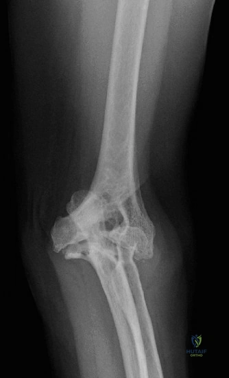
Describe the radiographic findings and outline your systematic approach to managing this patient.
Candidate: The radiographs show advanced rheumatoid elbow arthritis with joint space narrowing, marginal erosions of the trochlear notch and capitellum, and significant subchondral cystic changes. Given her functional impairment and failure of non-operative management, I would counsel the patient for a total elbow arthroplasty (TEA). I would assess her triceps function and bone stock pre-operatively, likely using a CT scan for templating.
Candidates often jump straight to "I would do a total elbow replacement" without mentioning the vital step of assessing the systemic health, skin quality, or the status of the ulnar nerve. Failing to address the "why" (failed conservative care) or missing the need for a formal assessment of the contralateral side and patient expectations will lower the score.
A structured response: 1. Radiographic Analysis: Describe the "RA Triad"—pannus-induced marginal erosions, generalized osteopenia, and joint space destruction. 2. Patient Evaluation: Mention the need for a multi-disciplinary review (rheumatology input for immunosuppression timing). 3. Surgical Rationale: Categorize the management into: Patient Factors (function/demands), Anatomical Factors (bone stock/ligamentous laxity), and Joint Factors (fixed flexion deformity/instability). 4. Technique: Mention the use of a semi-constrained linked prosthesis and the necessity of prophylactic ulnar nerve transposition, as it is standard of care in the RA elbow.
During your exposure for the total elbow arthroplasty, you are deciding on the triceps management. The patient has thin, friable skin and poor bone quality due to long-term steroid use. Explain your choice of triceps management and the implications for the ulnar nerve.
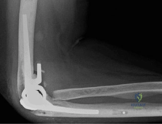
Candidate: I would consider an olecranon osteotomy because it provides the best exposure in cases where the bone is friable and visibility is paramount. For the ulnar nerve, I would perform a formal neurolysis and proceed with anterior submuscular transposition to protect it from tension during the postoperative phase.
Simply stating one technique is insufficient. The examiner wants to see you weigh the risks. A failing candidate will ignore the high risk of non-union of the olecranon in a patient with osteoporotic bone. You must discuss the trade-off: exposure versus the risk of hardware prominence and non-union.
Compare the techniques: The Bryan-Morrey (triceps-splitting) approach is more anatomical but offers less exposure. The olecranon osteotomy provides the best visualization for addressing severe deformity but carries a risk of non-union or hardware irritation. In an RA patient with friable tissue, I would choose based on the severity of the deformity. If I proceed with an osteotomy, I would use tension band wiring or a small plate, emphasizing the need for absolute stability to facilitate early protected range of motion. I would also mandate anterior submuscular transposition to avoid the "tethering effect" of the ulnar nerve against the medial side of the implant.
Post-operatively, the patient returns at 6 weeks complaining of persistent numbness in the ring and little finger and increasing pain. What is your differential, and how do you investigate this?
Candidate: The primary concern is ulnar nerve irritation or entrapment. My differential includes iatrogenic nerve injury during transposition, hematoma formation, or excessive scar tissue. I would perform a thorough neurological exam, potentially EMG/NCS if the symptoms don't resolve, and check for infection if there is any redness or warmth.
Failing to mention the most critical red flag: Infection. Any persistent pain and neurological changes after an arthroplasty must be presumed infected until proven otherwise.
State that the first priority is to exclude deep prosthetic joint infection (PJI). I would order inflammatory markers (CRP and ESR). If these are elevated, I would proceed to an aspiration of the joint. Regarding the ulnar nerve, I would assess if the nerve is firing or if there is a block. I would discuss the possibility of 'nerve tethering' at the site of transposition or local compression from hematoma. If infection is ruled out and the symptoms persist, I would consider surgical exploration to ensure the nerve is adequately transposed and free from fibrosis or hardware impingement.
