Moberg Advancement Flap: Comprehensive Guide to Surgical Technique & Outcomes for Thumb Reconstruction
Key Takeaway
The Moberg advancement flap is a foundational technique in reconstructive hand surgery. It restores sensate soft tissue coverage to full-thickness distal thumb pulp defects (typically 1-3 cm), preserving critical tactile sensation and functional grip/pinch. This local, innervated, vascularized flap is crucial for optimal thumb function after trauma.
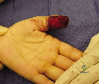
A 32-year-old carpenter presents following a power saw injury to the right thumb. Examination reveals a 2.5 cm full-thickness volar pulp defect with exposed distal phalanx. The IP joint is stable, but there is no distal sensation. The proper digital arteries and nerves appear clinically intact proximal to the defect. You are planning a Moberg advancement flap. Describe the anatomical basis for this flap and explain why it is preferred for this specific injury.
Candidate: The Moberg flap is a volar advancement flap. It is a bipedicled axial flap that includes both neurovascular bundles. Because the digital nerves are included, the flap provides sensate coverage. It is ideal for thumb defects up to 2-3 cm because it uses local tissue which is similar in texture and sensibility to the original pulp.
Failing to mention the "axial" nature of the flap or the reliance on the volar neurovascular bundles. Many candidates forget to mention the release of the fibrous septa, which is the most common reason for failure to achieve adequate advancement.
The Moberg flap is an axial bipedicled advancement flap. Its anatomical basis relies on the integrity of both proper digital neurovascular bundles which provide the vascular supply and sensation. The key to successful advancement is the meticulous division of the fibrous septa tethering the pulp to the flexor sheath. It is the gold standard here because it provides 'like-with-like' tissue, restores protective sensation—crucial for thumb function—and avoids the donor site morbidity of distant flaps.
During the procedure, you have mobilized the flap, but you are unable to achieve full coverage of the 2.5 cm defect without significant tension. How do you manage this intraoperatively?
Candidate: I would stop forcing the advancement to avoid necrosis. I would use a V-Y advancement technique or consider leaving a proximal defect and covering it with a split-thickness skin graft (STSG). I must ensure the flap is tension-free.
Suggesting a "unilateral pedicle division" to gain length without acknowledging the severe risk to flap viability. Also, failing to emphasize that the STSG is placed at the *proximal* donor site, not the defect site, to maximize the sensate pulp coverage.
Tension is the primary cause of Moberg flap necrosis. If maximal mobilization (including release of the fibrous septa and potentially the oblique retinacular ligament) is insufficient, I would utilize the 'V-Y' advancement or, more commonly, accept the advancement achievable and cover the resulting proximal volar defect with a full-thickness or split-thickness skin graft. This prevents distal flap tension while ensuring the distal thumb tip remains reconstructed with the original sensate, glabrous skin.
What are the contraindications to performing a Moberg advancement flap in the thumb?
Candidate: Contraindications include defects larger than 3 cm, damage to the digital neurovascular bundles, and severe crushing of the proximal volar skin.
Listing only one or two points. A senior candidate should categorize their answer (Defect, Vascular, Patient Factors) to show depth of knowledge.
I classify contraindications into three domains: 1. Defect Factors: Defects >3 cm or those involving extensive bone loss/unstable joints where local tissue is insufficient. 2. Vascular Factors: Damage to the proper digital neurovascular bundles proximal to the defect, or severe proximal crush injury/scarring that compromises the vascularity of the flap. 3. Patient/Clinical Factors: Active infection at the site or unrealistic expectations regarding sensory recovery and potential cold intolerance.
