Thumb CMC Joint Arthritis: Epidemiology, Anatomy, Biomechanics & Diagnosis Guide
Key Takeaway
Basal joint arthritis (thumb CMC joint OA) is a common degenerative condition affecting the thumb's base. Characterized by pain and weakness, its diagnosis relies on clinical examination and radiographic findings, often revealing subluxation. Key elements in its progression include anterior oblique ligament laxity and altered joint biomechanics under repetitive stress.
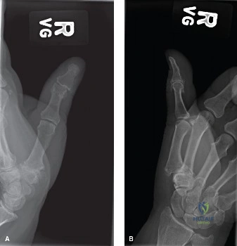
A 58-year-old female presents with a 6-month history of radial-sided thumb pain that is refractory to conservative management, including a thumb spica splint and two intra-articular corticosteroid injections. She reports weakness in key pinch and difficulty turning keys. On examination, you elicit a positive "grind test" and note a mild adduction contracture of the first metacarpal. You order the following radiographic series.
How would you classify this patient’s arthritis, and what are the specific clinical implications of this stage?
Candidate: This patient has Eaton-Littler Stage III or IV arthritis. She has joint space narrowing, osteophytes, and subluxation. Clinically, this explains her pain and weakness. I would recommend considering surgery because she has failed conservative management.
Candidates often fail to classify the stage precisely according to Eaton-Littler or neglect the "Z-deformity" associated with later stages. They also jump to "surgery" without differentiating whether the arthritis involves the STT joint, which changes the surgical plan from an isolated trapeziectomy to a potential partial wrist arthroplasty.
The candidate should state: "Based on the radiographs showing joint space narrowing, osteophytes (>2mm), and subluxation, this is Eaton-Littler Stage III. If the scaphotrapeziotrapezoid (STT) joint were involved, it would be Stage IV. At this stage, the pathogenesis involves the failure of the anterior oblique ligament, causing dorsoradial subluxation. Clinically, I would specifically look for an adduction contracture of the first metacarpal and compensatory MCP joint hyperextension, known as the Z-deformity. My management would transition to discussing surgical options, specifically trapeziectomy with ligament reconstruction and tendon interposition (LRTI), as she has failed non-operative treatment."
You have decided to proceed with a Trapeziectomy with LRTI. During the surgery, you are performing the suspensionplasty part of the procedure. Describe your steps for the suspensionplasty and explain how you determine the correct tension for the FCR graft.
Candidate: I would harvest a slip of the FCR tendon, keeping it attached proximally. I drill a hole in the base of the first metacarpal and pass the tendon through it. Then I wrap it around the APL tendon and suture it to itself to hold the thumb in place.
Vague descriptions of the drill hole location or failing to mention the importance of thumb positioning (abduction/pronation) during tensioning. Ignoring the "interposition" portion of the "LRTI" acronym is a major oversight.
The candidate must be precise: "I harvest a 1/3 to 1/2 width slip of the FCR tendon. I create a drill hole in the base of the first metacarpal from dorsal-radial to volar-ulnar. I pass the graft through the hole and tension it while the thumb is in 35-40 degrees of palmar abduction and slight pronation to restore the first web space. The tension must be sufficient to prevent proximal metacarpal subsidence but not so tight as to cause impingement against the scaphoid. The remaining graft is then rolled into a ball and sutured into the trapeziectomy space as an interposition spacer."
