Complex Intra-Articular Distal Radius Fracture: Case Study of Failed Volar Tilt Restoration
Key Takeaway
Failed closed reduction of an intra-articular distal radius fracture manifests as persistent dorsal angulation, inadequate volar tilt restoration, and articular step-off on post-reduction imaging. These issues confirm insufficient anatomical alignment, necessitating operative intervention for optimal functional outcomes and to prevent complications.
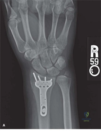
A 58-year-old female presents with a dorsally displaced, comminuted distal radius fracture after a FOOSH. A closed reduction attempt under sedation failed to restore acceptable alignment. You are reviewing the plain radiographs and the subsequent CT scan provided below.
Describe your systematic interpretation of these films and the specific pathological features that explain why the closed reduction failed.
Candidate: The X-ray shows a dorsally displaced fracture with shortening and loss of radial tilt. The CT is important because it shows the fracture involves the joint surface and the "die-punch" fragment of the lunate facet. The closed reduction likely failed because the fragment is rotated or comminuted, making it unstable in a cast.
Focusing only on the "broken pieces" without mentioning the 3D geometry. Failing to recognize that the primary reason for failure in these specific fractures is rotational malalignment (pronation of the distal fragment) and dorsal cortical comminution, which creates a mechanical block to reduction that simple traction cannot overcome.
Structure your answer: 1. Radiographic Parameters: Address radial height, radial inclination, and volar tilt. 2. CT Findings: Identify the specific intra-articular involvement (lunate/scaphoid facet) and the "die-punch" component. 3. The "Why": Explicitly state that the failure of closed reduction is due to the rotational component (pronation of the distal fragment) and the loss of the dorsal cortical buttress. Explain that ligamentotaxis alone cannot correct the rotational malalignment, which is the mechanical cause for the persistent dorsal angulation.
You have decided to proceed with an Open Reduction Internal Fixation (ORIF) using a volar locking plate. During your approach and reduction, what are the critical steps to ensure the volar tilt is restored and that you avoid long-term tendon complications?
Candidate: I would use the Henry approach, retracting the FCR. I would use a bone clamp to hold the fragment, restore the angle, and then put the plate on. I must make sure the plate is not too distal so it doesn't rub on the tendons.
Vague procedural steps. Candidates often miss the crucial "supination" maneuver of the distal fragment to correct the pronated malalignment, and fail to mention the "Watershed Line" as the anatomical landmark for safe plate placement.
Use a structured approach: 1. Reduction: Direct visualization after Pronator Quadratus (PQ) reflection. Emphasize the need to supinate the distal fragment while applying dorsal pressure to restore volar tilt. 2. Stability: Address the metaphyseal void with bone graft if necessary to prevent secondary collapse. 3. Hardware safety: Explicitly mention placing the plate proximal to the "Watershed Line" to prevent Flexor Pollicis Longus (FPL) tendon rupture. 4. Soft tissue: Mention meticulous repair of the PQ muscle to act as a barrier between the hardware and the flexor tendons.
