Complications of Distal Radius Fractures: Prevention, Biomechanics, and Surgical Management
Key Takeaway
Distal radius fractures present a broad spectrum of potential complications, ranging from malunion and hardware failure to tendon rupture and complex regional pain syndrome (CRPS). Overall complication rates vary from 6% to 80%. Successful management requires a profound understanding of wrist biomechanics, meticulous surgical technique—particularly regarding the watershed line during volar plating—and evidence-based postoperative protocols, including Vitamin C prophylaxis, to optimize patient outcomes and mitigate long-term morbidity.
Comprehensive Introduction and Patho-Epidemiology
The management of distal radius fractures has undergone a profound evolution over the past three decades, transitioning from traditional closed reduction and cast immobilization to sophisticated, anatomically contoured locked volar plating and fragment-specific osteosynthesis. Despite these technological and technical advancements, the distal radius remains a highly unforgiving anatomical region. The type, severity, and frequency of complications remain highly variable across reported series, presenting a continuous challenge to both novice and master orthopedic surgeons. In a comprehensive literature review, McKay et al. demonstrated that overall complication rates range dramatically from 6% to 80%, with rates of symptomatic posttraumatic arthritis spanning from 7% to 65% depending on the length of follow-up and the initial fracture severity.
Understanding the patho-epidemiology of these injuries is paramount for mitigating adverse outcomes. Distal radius fractures follow a bimodal epidemiological distribution. In younger cohorts, these injuries are typically the result of high-energy trauma—such as motor vehicle collisions or falls from significant heights—resulting in severe intraarticular comminution, profound soft tissue envelope compromise, and a high risk of acute complications such as compartment syndrome or acute carpal tunnel syndrome. Conversely, in the elderly, osteoporotic population, these fractures predominantly occur via low-energy mechanisms, such as a fall on an outstretched hand (FOOSH). In this demographic, the complications pivot toward hardware failure, loss of fixation in osteopenic bone, and delayed functional recovery secondary to profound stiffness.
The pathophysiology underlying complications in distal radius fractures is deeply intertwined with the biological response to trauma and iatrogenic intervention. The wrist is a densely packed anatomical crossroad of critical neurovascular structures and gliding tendon units, all encased within tight fascial compartments and retinacular sheaths. High-energy trauma incites a robust inflammatory cascade, leading to fibrinous exudate accumulation, subsequent fibroblast proliferation, and the formation of dense intra-articular and extra-articular adhesions. When surgical intervention is superimposed upon this traumatized tissue envelope, the risk of iatrogenic devascularization, tendon abrasion from prominent hardware, and nerve traction injuries increases exponentially.
As orthopedic surgeons, mitigating these inherent risks requires a profound understanding of carpal kinematics, meticulous surgical execution, and vigilant postoperative care. Complications can occur regardless of the management strategy chosen—operative or nonoperative—but the sequelae of specific complications can be significantly lessened by prompt, problem-specific intervention. The contemporary surgeon must not only be a master of osteosynthesis but also an astute diagnostician capable of recognizing the earliest signs of complex regional pain syndrome (CRPS), tendon attrition, or subtle loss of reduction before they evolve into irreversible clinical catastrophes.
Detailed Surgical Anatomy and Biomechanics
A rigorous comprehension of the osteology and ligamentous anatomy of the distal radius is the foundation upon which successful surgical reconstruction is built. The distal radius articulates with the scaphoid and lunate via two distinct, concave cartilaginous facets separated by a subtle interfacet ridge. Normal radiographic parameters are strictly defined and must be restored to optimize biomechanical function: radial inclination averages 22 to 24 degrees, palmar (volar) tilt is normally 10 to 12 degrees, radial height measures 11 to 12 mm relative to the ulnar head, and ulnar variance should be neutral. The sigmoid notch, located on the ulnar aspect of the distal radius, forms the distal radioulnar joint (DRUJ) and is critical for forearm pronation and supination.
The biomechanics of load transfer across the human wrist dictate that, in a state of neutral ulnar variance, approximately 80% of axial load is transmitted through the radiocarpal joint and 20% through the ulnocarpal joint. Even minor deviations from anatomic alignment drastically alter this distribution. For instance, radial shortening of a mere 2.5 mm shifts the load-bearing ratio to 60:40. This pathological load distribution rapidly leads to ulnocarpal impaction syndrome, characterized by degenerative attrition of the triangular fibrocartilage complex (TFCC), chondromalacia of the ulnar head and lunate, and debilitating ulnar-sided wrist pain. Similarly, a loss of volar tilt (dorsal angulation beyond neutral) shifts the center of rotation dorsally, increasing the load on the dorsal radiocarpal ligaments and predisposing the patient to midcarpal instability and dorsal intercalated segment instability (DISI).
The volar aspect of the distal radius features a critical anatomical landmark known as the "watershed line." First popularized by Orbay, this transverse ridge marks the most volar margin of the distal radius, where the robust volar radiocarpal ligaments originate. Proximal to this line lies the pronator fossa, a concave recess that safely accommodates volar plating systems. However, at and distal to the watershed line, the flexor pollicis longus (FPL) and the flexor digitorum profundus (FDP) tendons lie in intimate, unprotected contact with the bone. Hardware placed distal to this line, or plates that fail to perfectly contour to the watershed ridge, create a mechanical fulcrum that inevitably leads to flexor tendon tenosynovitis and subsequent attritional rupture.
Dorsally, the anatomy is equally unforgiving. The dorsal surface is convex and acts as a pulley system for the extensor tendons, which are organized into six distinct compartments by the extensor retinaculum. Lister's tubercle, a prominent bony landmark, separates the second compartment (extensor carpi radialis longus and brevis) from the third compartment (extensor pollicis longus). The EPL tendon takes an acute 45-degree turn around Lister's tubercle, making it highly susceptible to mechanical abrasion from dorsally penetrating screws. The vascular supply to the distal radius is primarily derived from the anterior interosseous artery and delicate capsular vessels; aggressive dorsal stripping during surgical exposure can disrupt this tenuous blood supply, contributing to the rare but catastrophic complication of avascular necrosis or nonunion.
Exhaustive Indications and Contraindications
The decision to proceed with operative management of a distal radius fracture requires a nuanced synthesis of radiographic parameters, patient-specific physiological demands, and a thorough risk-benefit analysis. Lafontaine's criteria historically defined instability, but modern indications have been refined by the American Academy of Orthopaedic Surgeons (AAOS) and contemporary literature. Operative intervention is generally indicated for fractures that cannot be reduced or maintained in an acceptable alignment via closed means. Specific radiographic indications include an intra-articular step-off or gap greater than 2 mm, radial shortening exceeding 3 mm, dorsal angulation greater than 10 degrees past neutral, or volar tilt exceeding 20 degrees.
Beyond radiographic metrics, patient-specific factors heavily influence the surgical calculus. High-energy mechanisms resulting in severe comminution, open fractures, or concomitant neurovascular compromise (e.g., acute carpal tunnel syndrome) mandate urgent surgical stabilization. Furthermore, the functional demands of the patient play a critical role; young, active individuals and manual laborers require near-anatomic restoration to prevent early-onset arthrosis and maintain grip strength. Conversely, in the low-demand, elderly patient with severe medical comorbidities, the functional detriment of a mild malunion may be vastly outweighed by the perioperative risks of general anesthesia and surgical morbidity.
Contraindications to operative management must be meticulously respected to avoid catastrophic outcomes. Absolute contraindications include active, untreated local soft tissue or bone infections, and patients who are medically unfit to tolerate anesthesia due to severe cardiopulmonary instability. Relative contraindications require careful surgeon judgment and often necessitate a modification of the surgical plan. For example, profound osteopenia or severe soft tissue compromise (e.g., massive fracture blisters, severe degloving) may preclude the use of immediate internal fixation with a volar locking plate. In such scenarios, temporary spanning external fixation or closed reduction and percutaneous pinning (CRPP) may be utilized as a bridge to definitive fixation or as the definitive treatment itself.
The following table delineates the comprehensive indications and contraindications for the operative management of distal radius fractures:
| Category | Operative Indications | Absolute Contraindications | Relative Contraindications |
|---|---|---|---|
| Radiographic | Intra-articular step-off > 2mm; Radial shortening > 3mm; Dorsal tilt > 10° | N/A | Severe osteopenia precluding screw purchase |
| Clinical/Anatomic | Open fractures; Concomitant carpal fractures; Polytrauma | Active local infection (e.g., cellulitis, osteomyelitis) | Massive soft tissue swelling/blistering |
| Neurological | Acute Carpal Tunnel Syndrome requiring release | N/A | Pre-existing severe peripheral neuropathy |
| Patient Factors | High functional demand; Failure of conservative cast management | Medically unfit for anesthesia (ASA IV/V) | Low functional demand elderly; Poor compliance |
Pre-Operative Planning, Templating, and Patient Positioning
Meticulous pre-operative planning is the cornerstone of successful distal radius fracture management and the primary defense against intraoperative surprises and subsequent complications. Standard radiographic evaluation must include true posteroanterior (PA), lateral, and oblique views of the wrist. However, for complex intra-articular fracture patterns, plain radiography is often insufficient. A non-contrast Computed Tomography (CT) scan with two-dimensional multiplanar reformats and three-dimensional surface rendering is highly recommended. CT imaging allows for the precise identification of critical fracture fragments—such as the dorsal ulnar "die-punch" fragment, the volar marginal lip of the lunate facet, and the degree of central articular impaction—which dictate the specific trajectory of fixation required.
Digital pre-operative templating is mandatory when utilizing modern volar locking plate systems. The surgeon must evaluate the size of the distal radius to select the appropriate plate width (e.g., standard, wide, or narrow) and anticipate the required screw lengths. Templating helps prevent the common error of selecting a plate that is too long, which can impinge on the diaphyseal bow of the radius, or a plate that is positioned too distally, encroaching upon the watershed line. Furthermore, in cases of profound metaphyseal comminution, the surgeon must plan for the potential use of bone graft substitutes (e.g., calcium phosphate cement or cancellous allograft) to provide structural support and prevent late secondary subsidence of the articular surface.
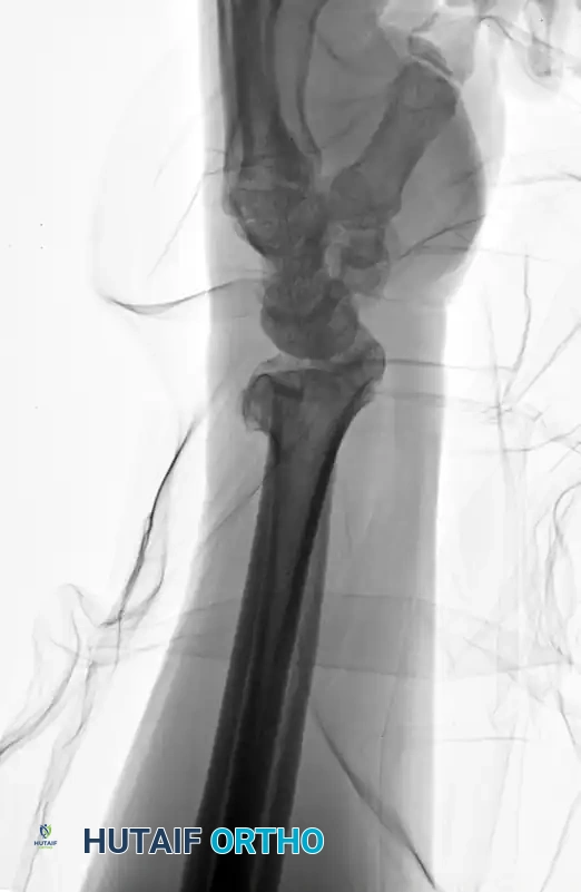
Preoperative AP radiograph demonstrating a highly comminuted, unstable distal radius fracture requiring meticulous preoperative templating.
Preoperative Lateral radiograph highlighting severe dorsal comminution and loss of volar tilt, necessitating structural restoration.
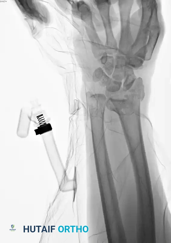
Patient positioning and operating room setup must be standardized to ensure seamless surgical flow and optimal fluoroscopic imaging. The patient is placed supine on the operating table with the operative extremity extended onto a radiolucent hand table. A well-padded pneumatic tourniquet is applied to the proximal arm. Following exsanguination with an Esmarch bandage, the tourniquet is inflated to 250 mmHg (or 100 mmHg above the patient's systolic blood pressure). The C-arm fluoroscopy unit should be positioned parallel to the hand table, entering either from the head of the bed or the contralateral side. This configuration allows the surgeon to obtain unobstructed AP, lateral, and crucial dorsal tangential views without breaking the sterile field or compromising the reduction.
The surgical team must also ensure that all necessary equipment is readily available before the incision is made. This includes not only the primary volar locking plate system but also fragment-specific fixation sets, variable-angle locking screws, K-wires for provisional fixation, and a mini-external fixator system in the event that internal fixation proves impossible due to extreme comminution or poor bone quality. A comprehensive "Plan B" is essential; the inability to pivot intraoperatively when faced with unexpected anatomical challenges is a primary driver of iatrogenic complications.
Step-by-Step Surgical Approach and Fixation Technique
The surgical execution of distal radius osteosynthesis demands a rigorous adherence to anatomical planes and biomechanical principles. The Modified Henry Approach is the gold standard for volar exposure. A longitudinal incision is made directly over the flexor carpi radialis (FCR) tendon, extending approximately 8 to 10 cm proximally from the distal wrist crease. The superficial fascia is incised, and the FCR tendon sheath is opened. The FCR tendon is meticulously retracted ulnarly. This specific retraction vector is critical; it protects the palmar cutaneous branch of the median nerve, which courses in the subcutaneous tissue immediately ulnar to the FCR. The deep fascial floor of the FCR sheath is then incised, exposing the deep compartment. The radial artery and its venae comitantes are identified and gently retracted radially, utilizing blunt retractors to avoid vasospasm or intimal injury.
Deep dissection reveals the pronator quadratus (PQ) muscle, which blankets the volar metaphysis of the distal radius. An L-shaped incision is made along the radial and distal borders of the PQ, leaving a small cuff of tissue for later repair. The PQ is elevated subperiosteally from radial to ulnar, exposing the fracture site. At this juncture, the fracture hematoma is thoroughly irrigated and debrided to allow for direct visualization of the articular and metaphyseal fragments. Reduction is achieved through a combination of longitudinal traction, volar translation, and direct manipulation of the fragments using a periosteal elevator or a dental pick. Once anatomic alignment is restored, provisional fixation is achieved using 0.045-inch or 0.062-inch K-wires strategically placed so as not to interfere with subsequent plate positioning.
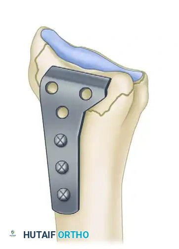
The L-plate provides a volar buttress to the volar rim of the lunate facet, yet allows fixation to the subcutaneous radial side of the proximal fragment, minimizing tendon abrasion.
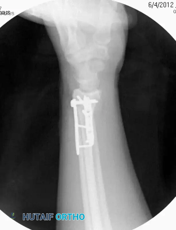
The application of the volar locking plate requires exact spatial awareness. The plate must be positioned strictly proximal to the watershed line to avert flexor tendon impingement. An oblong cortical screw is typically placed in the diaphyseal slot to allow for proximal-distal micro-adjustments. Once the plate position is verified fluoroscopically, the distal locking screws are inserted. These screws must provide subchondral support to the articular surface without penetrating the radiocarpal or DRUJ articulations. Crucially, a dorsal tangential fluoroscopic view (the "skyline" view) must be obtained to definitively rule out dorsal screw protrusion, which is the leading cause of iatrogenic extensor pollicis longus (EPL) rupture.
In scenarios involving severe, multi-fragmentary intra-articular comminution where a standard volar plate cannot adequately capture all fragments, fragment-specific fixation is mandated. This modular approach utilizes low-profile, wire-form plates, radial pin plates, and small-fragment screws to independently address the radial column, the intermediate (lunate facet) column, and the dorsal ulnar corner. By utilizing multiple smaller implants rather than a single bulky construct, the surgeon can achieve rigid, multi-planar osteosynthesis while minimizing soft tissue stripping. Following final fixation and fluoroscopic confirmation, the pronator quadratus is meticulously repaired over the hardware whenever possible, serving as a biological interpositional barrier between the plate and the traversing flexor tendons.
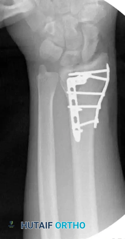
Postoperative AP radiograph demonstrating anatomic restoration of radial height and inclination utilizing fragment-specific fixation.
Postoperative Lateral radiograph confirming restoration of volar tilt and congruent radiocarpal articulation without dorsal screw penetration.
Complications, Incidence Rates, and Salvage Management
Despite optimal surgical technique, complications following distal radius fractures remain a formidable clinical reality. Hardware and tendon complications represent a significant source of iatrogenic morbidity, particularly in the era of locked volar plating. Flexor tendon tenosynovitis and subsequent rupture (most notably the FPL) occur in up to 12% of cases, driven by plate prominence distal to the watershed line. Extensor tendon ruptures, primarily the EPL, occur due to unrecognized dorsal screw penetration. Salvage management for tendon ruptures requires prompt hardware removal and tendon reconstruction, most commonly achieved via an extensor indicis proprius (EIP) to EPL tendon transfer, or an FDS to FPL transfer for volar injuries.
Neurological complications affect up to 17% of patients. Acute carpal tunnel syndrome (CTS) is a surgical emergency driven by fracture hematoma, severe displacement, or massive edema, requiring immediate carpal tunnel release alongside fracture stabilization. Delayed median neuropathy presents weeks to months post-injury and is often associated with malunion, exuberant callus formation, or prolonged immobilization in extreme flexion (the historical and dangerous Cotton-Loder position). If conservative measures fail, delayed surgical decompression and neurolysis are indicated. Iatrogenic injury to the superficial sensory branch of the radial nerve (SBRN) can occur during percutaneous pinning or external fixator application, leading to debilitating neuromas that may require surgical excision and burying of the nerve stump into local muscle.
Malunion remains the most frequent complication, with an incidence of approximately 17%, particularly following nonoperative management. Intra-articular step-offs greater than 2 mm are highly predictive of early-onset posttraumatic radiocarpal arthrosis. Extra-articular malunions (loss of radial height, dorsal tilt) lead to profound biomechanical derangements, including ulnocarpal impaction and midcarpal instability. Salvage of symptomatic malunions requires complex corrective osteotomies, utilizing structural bone graft and rigid internal fixation to restore anatomic parameters. If advanced arthrosis has already developed, salvage procedures such as total wrist arthrodesis, proximal row carpectomy (PRC), or partial wrist fusions (e.g., radioscapholunate fusion) become necessary to provide a painless, albeit stiff, wrist.
Complex Regional Pain Syndrome (CRPS) is a devastating neuro-inflammatory complication characterized by disproportionate pain, hyperalgesia, and sudomotor changes, occurring in up to 35% of susceptible patients. Prophylaxis is paramount; the administration of Vitamin C (500 mg daily for 50 days) has been shown to significantly reduce CRPS incidence. If CRPS develops, aggressive multidisciplinary management involving stress-loading hand therapy, neuropathic pain modulators (gabapentin), and sympathetic nerve blocks is critical. Nonunion of the distal radius is exceptionally rare (<1%), typically resulting from severe biological compromise (infection, massive devascularization) or mechanical failure, requiring revision osteosynthesis with autologous bone grafting.
| Complication | Incidence Rate | Primary Etiology / Risk Factor | Salvage / Management Strategy |
|---|---|---|---|
| Arthritis/Arthrosis | 7% – 65% | Intra-articular step-off >2mm; cartilage necrosis | NSAIDs, Injections; Salvage: Partial/Total Wrist Fusion |
| Tendon Rupture (FPL/EPL) | 0% – 12% | Plate distal to watershed line (FPL); Dorsal screw penetration (EPL) | Hardware removal; Tendon transfer (e.g., EIP to EPL) |
| Nerve Compression (CTS) | 0% – 17% | Acute hematoma; Malunion; Cotton-Loder positioning | Acute/Delayed Carpal Tunnel Release; Neurolysis |
| CRPS | 0.3% – 35% | Prolonged immobilization; Psychological factors | Prevention: Vitamin C; Treatment: Stress loading, blocks |
| Malunion | ~17% | Loss of reduction in cast; Inadequate surgical fixation | Corrective osteotomy; Ulnar shortening osteotomy |
| Delayed Union/Nonunion | 0.7% – 4% | Infection; Severe comminution; Devascularization | Revision rigid osteosynthesis with autologous bone graft |
Phased Post-Operative Rehabilitation Protocols
The ultimate objective of distal radius fracture management is not merely radiographic union, but the restoration of a painless, stable, and highly functional wrist. Achieving this goal requires a meticulously structured, phased postoperative rehabilitation protocol that balances the mechanical need for fracture stability with the biological imperative to prevent soft tissue adhesions and joint contractures. The frequency of complications, particularly profound stiffness and CRPS, is inversely related to the quality and timeliness of postoperative therapy.
Phase I (0-2 Weeks): The Protective Phase
Immediately following surgery, the wrist is immobilized in a bulky, non-constrictive compressive dressing reinforced with a removable volar splint. The primary goals during this acute phase are edema control, pain management, and the prevention of distal stiffness. Strict elevation of the extremity above the level of the heart is enforced. Patients are instructed to initiate immediate, aggressive active range of motion (AROM) of the fingers, thumb, elbow, and shoulder. Specifically, tendon gliding exercises (straight, hook, fist, and tabletop positions) are mandatory to prevent adhesions of the FPL and FDP tendons over the surgical site. Passive manipulation of the wrist is strictly contraindicated during this phase to protect the healing soft tissues and the provisional stages of osteosynthesis.
Phase II (2-6 Weeks): The Mobilization Phase
At the 10-to-14-day mark, sutures are removed, and the surgical incision is evaluated. If rigid internal fixation was achieved using a modern locked volar plate construct, the patient may safely transition out of continuous rigid immobilization. A custom-molded thermoplastic splint is fabricated, to be worn during sleep and high-risk activities, but removed for multiple daily exercise sessions. Under the guidance of a certified hand therapist, the patient begins gentle, gravity-eliminated AROM of the radiocarpal joint (flexion, extension) and the distal radioulnar joint (pronation, supination). Proprioceptive neuromuscular facilitation techniques are introduced to re-educate the stabilizing musculature of the forearm. Excessive passive stretching remains restricted to prevent hardware failure in osteopenic bone.
Phase III (6-12 Weeks): The Strengthening Phase
By 6 weeks postoperatively, clinical and radiographic evidence of bridging callus and early union is typically present. The protective splint is systematically weaned and discontinued. The rehabilitation focus shifts toward restoring end-range kinematics and building functional strength. Passive range of motion (PROM) exercises and joint mobilization techniques are introduced to address residual capsular contractures. Progressive resistive exercises using putty, hand dynamometers, and free weights are initiated to rebuild grip and pinch strength. If the patient exhibits recalcitrant stiffness, static progressive splinting or dynamic turnbuckle orthoses may be prescribed to provide a low-load, prolonged stretch to the contracted tissues.
Phase IV (12+ Weeks): Return to High Demand
Beyond 12 weeks, the bone is generally considered structurally sound enough to withstand physiologic loading without risk of displacement. Patients are cleared for a graduated return to heavy manual labor, contact sports, and high-impact activities. However, it is imperative to counsel patients that maximal medical improvement following a severe distal radius fracture may take up to 12 to 18 months. Intermittent aching, weather-related discomfort, and minor deficits in terminal flexion or supination are common and should be managed with realistic expectation setting during the preoperative and early postoperative periods.
Summary of Landmark Literature and Clinical Guidelines
The contemporary management of distal radius fractures is heavily guided by an extensive body of high-quality, peer-reviewed literature and formalized clinical practice guidelines. The American Academy of Orthopaedic Surgeons (AAOS) has published rigorously vetted guidelines that serve as the benchmark for evidence-based practice. Among their strongest recommendations is the prophylactic administration of Vitamin C to mitigate the risk of Complex Regional Pain Syndrome, a practice supported by multiple randomized controlled trials demonstrating a significant reduction in CRPS incidence when 500 mg is administered daily for 50 days post-injury. Furthermore, the AAOS strongly supports the use of rigid immobilization (whether via cast or internal fixation) to prevent loss of reduction, while noting that the choice between operative and nonoperative management in the elderly population must be highly individualized.
The WRIST trial (Wrist and Radius Injury Surgical Trial), a landmark multicenter randomized controlled study, provided critical insights into the management of older adults (aged 60 and above) with unstable distal radius fractures. The trial compared outcomes across three operative modalities: volar locking plates, closed reduction and percutaneous pinning (CRPP), and external fixation. Surprisingly, the study concluded that at 12 and 24 months postoperatively, there were no clinically significant differences in functional outcomes (as measured by the Michigan Hand Outcomes Questionnaire) among the three surgical groups. This pivotal data underscores the principle that in older, lower-demand patients, the specific hardware utilized is less critical than achieving an acceptable reduction and initiating early, aggressive rehabilitation.
The biomechanical and clinical foundations of articular congruity were established by the seminal works of Jupiter, Fernandez, and Knirk. Their long-term follow-up studies definitively established the "2-millimeter rule," demonstrating that residual intra-articular
