
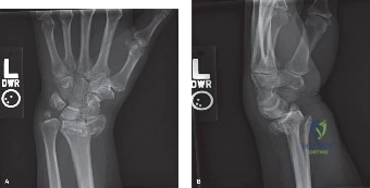
A 72-year-old female presents to the ED following a low-energy fall onto an outstretched hand. She is non-diabetic and otherwise fit. Radiographs are provided below. Describe your clinical assessment and your decision-making process for this patient.
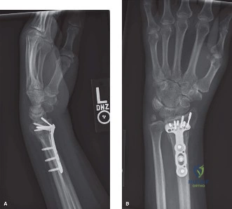
Candidate: I would perform a thorough neurovascular assessment, checking median nerve function specifically. I would evaluate the radiographs to assess radial height, inclination, and volar tilt. Given her age and the fracture pattern, I would determine if this is stable or unstable. If it meets criteria for non-operative management, I would attempt a closed reduction and immobilization.
The candidate focuses only on the radius. They fail to mention the contralateral wrist as a reference, ignore the status of the DRUJ, and fail to provide a structured "Indications vs. Contraindications" framework. They also miss the importance of asking about functional demand vs. chronological age.
I would approach this systematically: 1. Clinical: Assess neurovascular status (median nerve), soft tissue, and baseline function. 2. Imaging: Compare with the contralateral wrist to establish anatomical baseline. 3. Classification: Utilize AO/OTA classification. 4. Decision: If the fracture is stable (or reducible to acceptable parameters: radial inclination >15°, volar tilt neutral, shortening <3-5mm, and intra-articular step <1-2mm), I would proceed with non-operative management. If the criteria for instability are met—specifically intra-articular gap, dorsal comminution, or persistent DRUJ instability—I would advocate for volar locking plate fixation to ensure early mobilization and minimize CRPS risk.
During the volar Henry approach to the radius, you encounter the pronator quadratus. How do you handle this structure, and why is this technique relevant to long-term outcomes?
Candidate: I would use an L-shaped incision to reflect the pronator quadratus from radial to ulnar. I would then repair it at the end of the procedure. This is important to cover the plate and protect the tendons.
The candidate ignores the vascularity of the muscle and fails to mention its role in DRUJ stability. They also miss the specific benefit regarding the reduction of tendon irritation, which is a known cause of FPL rupture.
I perform an L-shaped capsulotomy and release the pronator quadratus from its radial attachment, reflecting it ulnar-wards. This creates a vascularized soft tissue envelope. Repairing it is critical because it acts as a mechanical barrier between the hardware and the flexor tendons (specifically the FPL), significantly reducing the risk of iatrogenic tendon rupture. Furthermore, it contributes to the vascular supply of the distal radius and provides ancillary stability to the DRUJ.
You have successfully fixed a distal radius fracture. What is your strategy for the prevention of Complex Regional Pain Syndrome (CRPS) in the perioperative period?
Candidate: I would ensure good pain control, early mobilization, and advise the patient to keep the hand elevated. Some people suggest using Vitamin C.
The candidate is too vague. They fail to mention the standardized prophylactic dosage of Vitamin C (500mg/50 days) and fail to categorize the approach into pre-, intra-, and post-operative stages.
CRPS prevention must be a multimodal, phase-specific strategy. 1. Pre-op: Prophylactic Vitamin C (500mg daily for 50 days) and identification of high-risk patients. 2. Intra-op: Meticulous tissue handling, minimizing tourniquet time, and utilizing regional anesthetic blocks for superior post-op analgesia. 3. Post-op: Aggressive edema management (elevation), early controlled active motion of digits to maintain motor cortex input, and patient education to mitigate anxiety. Early involvement of hand therapy is essential.
Detailed Chapters & Topics
Dive deeper into specialized chapters regarding distal-radius-fracture