Part of the Master Guide

Comprehensive Guide to Humerus & Elbow: Surgical Anatomy, Biomechanics, & Pathologies

Detailed Clinical Diagnosis of Lateral Epicondylitis: Patient Case & Findings

17 Apr 2026 18 min read 96 Views
Illustration of elbow cases lateral - Dr. Mohammed Hutaif

Key Takeaway

Lateral epicondylitis, or tennis elbow, is diagnosed through a detailed patient history of lateral elbow pain, often exacerbated by wrist extension. Key clinical findings include exquisite tenderness over the ECRB origin, and positive special tests like Cozen's, Mill's, and Maudsley's, which reproduce pain with resisted wrist/finger extension. Imaging typically rules out other pathology.
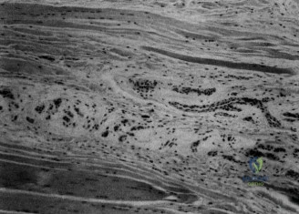
Patient Presentation and History

Demographic Profile and Occupational Biomechanics

A 48-year-old right-hand dominant male presented to the orthopedic clinic with a chief complaint of chronic, progressive right elbow pain. The patient is an avid recreational tennis player, participating in matches three to four times per week, and works as a graphic designer, a profession requiring prolonged, static wrist extension and repetitive micro-movements of the digits. The convergence of these two activities places an immense biomechanical load on the extensor origin of the lateral elbow. The kinematics of the tennis one-handed backhand stroke, particularly when executed with suboptimal technique leading to a "leading wrist" rather than a locked wrist, transmits extreme eccentric forces directly through the extensor carpi radialis brevis (ECRB) musculotendinous unit. Furthermore, the sustained isometric contraction required for mouse and keyboard manipulation creates a baseline of inadequate tissue perfusion and chronic mechanical stress, predisposing the tendinous origin to microvascular compromise and subsequent degenerative changes.

Chronology of Symptoms and Prior Interventions

His symptoms began approximately nine months prior to presentation, insidious in onset, and gradually worsening over time. Initially, the pain was primarily associated with the eccentric loading phases of his tennis backhand and heavy lifting tasks. Over the last three months, the pain has transitioned into a constant state, interfering with activities of daily living such as gripping objects, typing, and the simple act of shaking hands. He describes the pain as a constant, deep, dull ache localized to the anterior aspect of the lateral epicondyle, with occasional radiation down the dorsal forearm towards the radiocarpal joint. He denies any acute traumatic event, subjective feelings of instability, mechanical locking, or overt neurological symptoms such as distal numbness or paresthesias.

The patient reports having undergone an exhaustive course of conservative management initiated by his primary care physician. This regimen included strict activity modification, a prolonged course of oral non-steroidal anti-inflammatory drugs (ibuprofen 600mg TID prn), the utilization of a counterforce orthosis designed to dissipate tensile forces away from the epicondylar origin, and a structured physical therapy program. The physical therapy focused heavily on eccentric loading protocols for the wrist extensors, which is the current gold standard for non-operative tendinopathy management. These measures provided only transient, minimal relief, with symptoms invariably recurring upon resumption of baseline activities.

Crucially, the patient received a single corticosteroid injection into the peritendinous region of the lateral epicondyle four months prior to this consultation. While this provided profound symptomatic relief for approximately two weeks, the pain returned to baseline severity shortly thereafter. From a pathophysiological standpoint, while corticosteroids provide potent short-term anti-inflammatory effects, they are known to be cytotoxic to tenocytes, inhibit collagen synthesis, and can accelerate the underlying angiofibroblastic degeneration if utilized repeatedly. Relevant medical history includes well-controlled essential hypertension and hyperlipidemia. He is otherwise healthy, a non-smoker, and reports occasional alcohol consumption. There is no prior history of elbow trauma, upper extremity fractures, or previous surgical interventions. His primary objective is a return to pain-free athletic participation and occupational duties.

Clinical Examination Findings

A comprehensive, systematic examination of the right upper extremity was performed to isolate the precise anatomical pain generator and rule out concurrent pathology.

Visual Inspection and Palpation Metrics

Visual inspection of the right elbow revealed no overt effusion, erythema, ecchymosis, or obvious osseous deformity of the radiocapitellar or ulnohumeral joints. There was no clinically detectable muscle atrophy in the extensor wad or distal forearm musculature when compared to the contralateral extremity. The cutaneous tissues overlying the lateral epicondyle appeared normal, with no dermal atrophy or hypopigmentation indicative of adverse reactions from the prior corticosteroid injection.

Palpation was meticulously performed to differentiate between the various structures of the lateral compartment. Exquisite point tenderness was elicited directly over the origin of the extensor carpi radialis brevis tendon, situated approximately one to two centimeters distal and slightly anterior to the prominence of the lateral epicondyle. Pain was maximal with deep, sustained palpation over this specific footprint. Conversely, no significant tenderness was noted over the common extensor origin (extensor digitorum communis), the anconeus, or the supinator muscle belly.

Ligamentous assessment revealed that the lateral ulnar collateral ligament and the radial collateral ligament were completely stable to palpation and applied varus stress testing, effectively ruling out posterolateral rotatory instability. The olecranon, medial epicondyle, and common flexor origin were non-tender. The radial head was non-tender and translated smoothly through the sigmoid notch during pronosupination. The capitellum was similarly non-tender.

Range of Motion and Provocative Maneuvers

Active and passive ranges of motion of the ulnohumeral and radiocapitellar joints were full and pain-free, demonstrating flexion from 0 to 145 degrees, extension to 0 degrees, supination to 85 degrees, and pronation to 80 degrees. The absence of a flexion contracture or terminal extension block suggests the absence of significant intra-articular pathology or advanced radiocapitellar osteoarthritis.

Provocative maneuvers designed to selectively load the ECRB consistently reproduced the patient's chief complaint:
* Cozen's Test (Resisted Wrist Extension): Severe, sharp pain was reported over the lateral epicondyle when the patient actively extended the wrist against resistance. This was most pronounced with the elbow in full extension and the forearm in maximal pronation, a position that places the ECRB under maximal tension and simulates the biomechanics of a tennis backhand stroke.
* Maudsley's Test (Resisted Middle Finger Extension): Significant pain was elicited over the lateral epicondyle during resisted extension of the third digit. This test is highly specific for ECRB pathology, as the ECRB tendon inserts at the base of the third metacarpal, and isolated resistance effectively tensions this specific musculotendinous unit independent of the extensor digitorum communis.
* Mill's Test (Passive Wrist Flexion): Passive maximal flexion of the wrist with the elbow in full extension and forearm pronated elicited a stretching pain over the lateral epicondyle, further confirming the presence of an extensor tendinopathy.
* Dynamometry and Grip Testing: Handheld dynamometry revealed a significant disparity in grip strength compared to the contralateral side. Furthermore, the act of forceful gripping reproduced the lateral elbow pain. This occurs because the wrist extensors must contract isometrically during gripping to stabilize the wrist in slight extension, thereby preventing the flexors from actively insufficiating.

Neurological and Kinetic Chain Assessment

A thorough neurological examination is paramount to exclude compressive neuropathies. There was no tenderness or Tinel's sign elicited over the radial nerve in the antecubital fossa or over the superficial sensory branch of the radial nerve at the wrist. Crucially, deep palpation over the course of the posterior interosseous nerve within the Arcade of Frohse and the supinator muscle belly was completely non-tender, significantly lowering the clinical probability of radial tunnel syndrome. The ulnar nerve was stable within the cubital tunnel with a negative Tinel's sign and no subluxation during dynamic flexion.

A kinetic chain assessment of the ipsilateral shoulder and cervical spine was performed. Cervical spine range of motion was full, and Spurling's maneuver was negative, ruling out a C6 radiculopathy which can occasionally refer pain to the lateral elbow. Scapular dyskinesia was not observed, and rotator cuff strength was symmetric, indicating that proximal kinetic chain deficits were not the primary drivers of the distal overload in this particular patient.

Imaging and Diagnostics

While the diagnosis of lateral epicondylitis is primarily clinical, advanced imaging modalities are critical in chronic, recalcitrant cases to confirm the extent of tendinosis, evaluate for partial or full-thickness macroscopic tearing, and systematically rule out intra-articular pathology prior to surgical intervention.

Radiographic Evaluation

Standard orthogonal radiography of the right elbow, including anteroposterior, true lateral, and radiocapitellar oblique projections, was obtained. The osseous structures demonstrated normal morphology and trabecular patterns. There was no evidence of acute fracture, periosteal reaction, or osteochondral defects. The radiocapitellar and ulnohumeral joint spaces were well-preserved, with no marginal osteophytosis or subchondral sclerosis that would suggest degenerative joint disease. Notably, there were no dystrophic calcifications visualized within the common extensor origin, a finding that can sometimes be seen in chronic tendinopathy or calcific tendinitis, and which would alter the surgical approach regarding the necessity of thorough debridement of calcific deposits.

Advanced Imaging Modalities

Given the failure of prolonged conservative management and the duration of symptoms exceeding nine months, Magnetic Resonance Imaging without intravenous contrast was indicated to precisely delineate the soft tissue architecture.

Image

The MRI protocol included fluid-sensitive sequences (T2-weighted with fat suppression and STIR) as well as T1-weighted anatomical sequences in the coronal, sagittal, and axial planes. The imaging confirmed the clinical diagnosis, revealing classic signs of severe, chronic tendinosis isolated to the ECRB origin.

Specifically, the coronal T2-weighted fat-suppressed images demonstrated a focal region of marked hyperintensity within the deep fibers of the ECRB origin, just distal to its attachment on the lateral epicondyle. This intermediate-to-high signal intensity replaces the normally low-signal, tightly packed collagen bundles of the healthy tendon, representing the accumulation of mucoid ground substance, extracellular fluid, and angiofibroblastic hyperplasia. Furthermore, there was evidence of a high-grade partial-thickness interstitial tear extending through approximately fifty percent of the tendon footprint, characterized by fluid signal bridging the tendon fibers. The overlying extensor digitorum communis aponeurosis remained intact, providing an anatomical explanation for the lack of gross functional deficit despite the severe pain.

The lateral ulnar collateral ligament was traced from its origin on the lateral epicondyle to its insertion on the supinator crest of the ulna and was found to be continuous and of normal low signal intensity. The radiocapitellar joint showed no evidence of an enlarged synovial plica, and the capitellar cartilage was pristine, definitively ruling out osteochondritis dissecans. The posterior interosseous nerve was visualized coursing through the supinator without any surrounding edema or space-occupying lesions.

Differential Diagnosis

The presentation of lateral elbow pain requires a meticulous differentiation between several distinct pathologies. The following diagnostic matrix outlines the primary competing diagnoses.

Diagnostic Matrix

Pathology Primary Pain Location Key Provocative Exam Findings Distinguishing Imaging Characteristics Pathophysiologic Mechanism
Lateral Epicondylitis Anterior/distal to lateral epicondyle (ECRB origin) Positive Cozen's, Maudsley's, and Mill's tests. Pain with resisted wrist/long finger extension. T2 hyperintensity/tearing at ECRB origin on MRI. Normal radiocapitellar joint. Angiofibroblastic tendinosis due to repetitive microtrauma and failed healing response.
Radial Tunnel Syndrome 3-4 cm distal to lateral epicondyle, over mobile wad Tenderness over supinator. Pain with resisted forearm supination. Positive middle finger test (sometimes). Often normal. MRI may show denervation edema in PIN distribution (rarely). Compression of the Posterior Interosseous Nerve (PIN) at the Arcade of Frohse.
Radiocapitellar Plica Posterolateral joint line Pain with terminal extension and pronation. Mechanical catching or snapping. Thickened synovial fold (>3mm) in the radiocapitellar joint on MRI. Hypertrophy of the posterolateral synovial fringe causing mechanical impingement.
Posterolateral Rotatory Instability Lateral elbow, vague instability Positive lateral pivot-shift test. Positive chair test. Apprehension with supination/extension. Disruption, attenuation, or tearing of the Lateral Ulnar Collateral Ligament (LUCL). Traumatic or iatrogenic insufficiency of the LUCL leading to radial head subluxation.
Capitellar Osteochondritis Dissecans Deep radiocapitellar joint Pain with valgus loading. Crepitus, mechanical locking, loss of extension. Subchondral radiolucency, loose bodies, or focal cartilage defects on MRI/CT. Focal ischemic necrosis of subchondral bone, typically in adolescent throwing athletes.

Competing Pathologies

Radial Tunnel Syndrome (RTS): This is the most critical differential to exclude, as it frequently coexists with or mimics lateral epicondylitis. RTS is a compressive neuropathy of the posterior interosseous nerve, most commonly at the proximal edge of the superficial head of the supinator (the Arcade of Frohse). Clinically, the pain in RTS is typically located further distal than in lateral epicondylitis, approximately three to four centimeters distal to the epicondyle within the mobile wad of Henry. Provocative testing involves resisted supination with the elbow extended, which actively tightens the supinator fascia over the nerve. In this patient, the absence of distal tenderness and negative provocative nerve tests effectively ruled out RTS.

Radiocapitellar Plica Syndrome: A hypertrophic synovial plica can cause localized lateral elbow pain, but this is typically characterized by mechanical symptoms such as snapping, catching, or a painful arc of motion during terminal extension and pronation. The pain is usually localized to the posterolateral joint line rather than the ECRB origin. The patient's lack of mechanical symptoms and normal intra-articular MRI findings excluded this pathology.

Posterolateral Rotatory Instability (PLRI): PLRI results from insufficiency of the lateral ulnar collateral ligament, often secondary to prior trauma, multiple corticosteroid injections, or previous overly aggressive surgical debridement of the lateral elbow. Patients typically present with symptoms of instability, apprehension, or a sense of the elbow "giving way" when pushing up from a chair. The patient's stable varus stress test, negative history of instability, and intact LUCL on MRI ruled out PLRI.

Surgical Decision Making and Classification

Histopathologic Considerations

To understand the rationale for surgical intervention, one must first understand the true histopathology of the disease. The term "epicondylitis" is a misnomer, as acute inflammatory cells (macrophages, lymphocytes, neutrophils) are conspicuously absent in chronic cases. The seminal work by Nirschl and Pettrone demonstrated that the underlying pathology is not inflammatory, but rather a degenerative process termed angiofibroblastic tendinosis.

This process is characterized by a failed healing response within the ECRB tendon. Macroscopically, the tissue appears gray, friable, and edematous. Microscopically, there is a disorganized proliferation of immature fibroblasts, an ingrowth of non-functional, chaotic microvasculature (neovascularization), and a disruption of the normal parallel orientation of type I collagen fibers, which are replaced by mechanically inferior type III collagen. Furthermore, there is an upregulation of nociceptive neurotransmitters such as Substance P and Calcitonin Gene-Related Peptide (CGRP) within the neovessels, directly contributing to the severe pain profile. The structural integrity of the tendon is compromised, leading to the micro-tearing and partial interstitial tearing observed on this patient's MRI.

Indications for Operative Intervention

Surgical intervention for lateral epicondylitis is rarely the first line of treatment. The vast majority of patients (80-90%) will respond to a comprehensive, prolonged course of non-operative management. However, operative intervention is strongly indicated in this specific patient based on the following criteria:
1. Duration of Symptoms: The patient has experienced debilitating symptoms for over nine months, well beyond the typical six-month threshold where spontaneous resolution becomes statistically unlikely.
2. Failure of Comprehensive Conservative Care: The patient has exhausted physical therapy (including eccentric loading), bracing, NSAIDs, and activity modification without sustained benefit.
3. Detrimental Prior Interventions: The prior corticosteroid injection, while temporarily palliative, likely contributed to further local tissue atrophy and inhibition of fibroblastic collagen synthesis, worsening the underlying tendinosis.
4. MRI Confirmation: Advanced imaging confirms a high-grade structural defect (partial tearing) and severe tendinosis of the ECRB origin, indicating a macroscopic failure of the tissue that is unlikely to remodel without surgical debridement.
5. Quality of Life Impact: The pathology is significantly impairing both his occupational duties as a graphic designer and his recreational athletic pursuits.

The surgical strategy selected for this patient is an open debridement of the ECRB origin with decortication of the lateral epicondyle (the classic Nirschl procedure). While arthroscopic and percutaneous techniques exist and offer theoretically faster initial recovery times, the open approach remains the gold standard. It allows for direct, macroscopic visualization of the pathologic tissue, precise excision of the angiofibroblastic lesion without violating the lateral ulnar collateral ligament, and secure repair of the overlying extensor aponeurosis. The literature demonstrates that open, arthroscopic, and percutaneous techniques yield essentially equivalent long-term functional outcomes, making the open approach highly reliable and safe in experienced hands.

Surgical Technique and Intervention

Anesthesia and Patient Positioning

The patient is taken to the operating theater and placed in the supine position on the operating table. Anesthesia is achieved via a regional supraclavicular brachial plexus block utilizing a long-acting local anesthetic (e.g., 0.5% Ropivacaine) combined with monitored anesthesia care (MAC) for patient comfort. This multimodal approach ensures excellent intraoperative operating conditions and profound postoperative analgesia.

A non-sterile pneumatic tourniquet is applied to the proximal brachium over soft padding. The right upper extremity is prepped and draped in the standard sterile orthopedic fashion. The arm is positioned on a radiolucent hand table, allowing for free mobility of the elbow and wrist during the procedure. The arm is exsanguinated using an Esmarch bandage, and the tourniquet is inflated to 250 mmHg to ensure a bloodless surgical field, which is critical for identifying the subtle tissue planes between the healthy and pathologic tendon fibers.

Surgical Approach and Deep Dissection

A longitudinal incision, approximately three to four centimeters in length, is made centered just anterior to the tip of the lateral epicondyle and extending distally over the extensor wad. The incision is carefully planned to follow the Langer lines where possible to optimize cosmetic healing.

Subcutaneous dissection is carried down through the adipose tissue. Meticulous hemostasis is maintained using bipolar electrocautery. Care is taken during this superficial dissection to identify and protect any crossing branches of the posterior antebrachial cutaneous nerve, retraction of which is performed gently using blunt Senn retractors.

The deep fascia overlying the extensor digitorum communis (EDC) is identified. A longitudinal incision is made directly in line with the skin incision through the EDC aponeurosis. The EDC fibers are then bluntly split longitudinally, revealing the underlying origin of the extensor carpi radialis brevis. The ECRB is anatomically distinct; it lies deep and slightly anterior to the EDC and originates from the lateral epicondyle, the radial collateral ligament, and the adjacent intermuscular septum.

Tendon Debridement and Epicondylar Decortication

Upon exposure of the ECRB origin, the macroscopic pathology is immediately evident. In contrast to the glistening, white, organized collagen of the overlying EDC, the pathological ECRB tissue (the Nirschl lesion) appears dull, gray, edematous, and friable.

Using a #15 blade scalpel, an elliptical excision of the diseased ECRB tissue is performed. The debridement must be aggressive enough to remove all angiofibroblastic tissue, extending down to the footprint on the anterior aspect of the lateral epicondyle. It is imperative during this deep dissection to stay anterior to the equator of the radiocapitellar joint to absolutely avoid iatrogenic injury to the lateral ulnar collateral ligament, which originates slightly posterior and distal to the ECRB footprint.

Once the pathological soft tissue is completely excised, the cortical bone of the lateral epicondyle is exposed. The cortex in this region is often sclerotic due to chronic traction and failed healing. To stimulate a robust, biologically active healing response, the footprint is decorticated. This is achieved using a 2.0mm drill bit or a surgical awl to create multiple vascular access channels (microfracture) into the underlying cancellous bone marrow. The egress of marrow elements, including mesenchymal stem cells and growth factors, into the operative bed will facilitate the formation of a healthy fibrocartilaginous scar capable of anchoring the remaining tendon fibers.

Following decortication, the surgical site is thoroughly irrigated with sterile normal saline to remove any bone debris. The tourniquet is then deflated, and meticulous hemostasis is achieved. The remaining healthy edges of the ECRB and the overlying EDC aponeurosis are re-approximated and repaired in a side-to-side fashion using running or interrupted #1 Vicryl sutures. This restores the structural tension of the extensor wad. The subcutaneous layer is closed with 2-0 Vicryl, and the skin is approximated using a running subcuticular 3-0 Monocryl suture. A sterile, non-adherent dressing is applied, followed by a well-padded posterior splint with the elbow immobilized in 90 degrees of flexion and the wrist in slight extension (20 degrees) to offload the repair.

Post Operative Protocol and Rehabilitation

The postoperative rehabilitation protocol is meticulously phased to protect the surgical repair while progressively restoring functional mobility and strength. The timeline is dictated by the biological phases of tissue healing.

Immediate Postoperative Phase

Phase I (Weeks 0-2): Protection and Wound Healing
The primary goal during the initial two weeks is to protect the surgical repair, manage postoperative edema, and ensure optimal wound healing. The patient remains strictly immobilized in the posterior splint applied in the operating room. Elevation and cryotherapy are highly encouraged to mitigate swelling. The patient is instructed to perform active range of motion of the shoulder and digits to prevent stiffness and promote venous return, but active wrist extension and forearm pronation are strictly prohibited. At the two-week mark, the patient returns to the clinic for splint removal, wound inspection, and suture removal if necessary.

Intermediate and Advanced Rehabilitation

Phase II (Weeks 2-6): Restoration of Mobility
Upon removal of the splint, the patient is transitioned into a removable hinged elbow brace or a simple counterforce strap, depending on patient compliance and comfort. Physical therapy is formally initiated. The focus shifts to restoring full active and passive range of motion of the elbow and wrist. Gentle isometric strengthening of the wrist extensors and flexors is introduced in pain-free arcs of motion. Supination and pronation exercises are advanced. The patient is cautioned against lifting objects heavier than a coffee cup and must avoid combined elbow extension and wrist flexion, which places maximal tensile stress on the healing ECRB footprint.

Phase III (Weeks 6-12): Concentric and Eccentric Strengthening
As fibroblastic proliferation transitions into the remodeling phase, the mechanical demands on the tissue must be increased to stimulate proper collagen alignment. Concentric strengthening of the forearm musculature is progressed. Crucially, eccentric loading protocols for the wrist extensors are heavily emphasized during this phase. Eccentric exercise has been biomechanically proven to optimize tendon remodeling and increase the load-to-failure strength of the musculotendinous unit. Kinetic chain integration begins, incorporating shoulder stabilization and core strengthening exercises to ensure that proximal deficits do not lead to distal overload upon return to activity.

Phase IV (Months 3-6): Return to Sport and Occupational Simulation
In the final phase of rehabilitation, the focus is on sport-specific and occupation-specific functional training. For this patient, this involves a gradual return to typing and graphic design tasks, implementing strict ergonomic modifications (e.g., vertical mouse, proper keyboard height)


Dr. Mohammed Hutaif
Medically Verified Content by
Prof. Dr. Mohammed Hutaif
Consultant Orthopedic & Spine Surgeon
Chapter Index