Operative Management of Elbow Contractures and Advanced Rotator Cuff Pathology
Key Takeaway
The operative management of elbow contractures requires meticulous surgical technique to restore functional range of motion while preserving joint stability. The Morrey technique utilizes a lateral approach, allowing comprehensive anterior and posterior capsulectomies, triceps tenolysis, and osteophyte debridement. Concurrently, understanding the biomechanics of shoulder impingement and rotator cuff pathology is essential for upper extremity surgeons. This guide details the step-by-step surgical execution, indications, and postoperative protocols for optimizing patient outcomes in complex upper limb reconstruction.
Comprehensive Introduction and Patho-Epidemiology
The management of complex upper extremity pathology demands an uncompromising and profound understanding of both biomechanics and surgical anatomy. This masterclass chapter bridges two critical, highly specialized domains of upper limb orthopaedics: the surgical release of the stiff elbow and the advanced management of rotator cuff disease. These two distinct yet mechanically related areas represent some of the most challenging clinical scenarios encountered by the orthopaedic surgeon, requiring a synthesis of meticulous soft-tissue handling, precise osseous debridement, and an advanced understanding of dynamic joint kinematics. The upper extremity functions as a highly coordinated kinetic chain; thus, profound deficits in either the elbow or the glenohumeral joint significantly compromise the spatial positioning and functional utility of the hand.
Elbow contractures, whether post-traumatic, degenerative, or congenital, severely limit the functional arc of motion. The seminal biomechanical studies by Morrey et al. established that the functional arc required for most activities of daily living (ADLs) encompasses 30 to 130 degrees of flexion, with 50 degrees of pronation and 50 degrees of supination. Patho-epidemiologically, post-traumatic arthrofibrosis is the most common etiology, often following distal humerus fractures, terrible triad injuries, or prolonged immobilization. The pathogenesis involves an intense fibroproliferative cascade, leading to a thickened, non-compliant anterior and posterior capsule. Furthermore, heterotopic ossification (HO) frequently complicates neurotrauma, burns, and direct high-energy elbow trauma, creating absolute mechanical blocks that require precise surgical excision once the woven bone has fully matured into lamellar bone.
Concurrently, shoulder pathology—ranging from subacromial impingement to massive, irreparable rotator cuff tears—requires a highly nuanced, evidence-based approach rooted in tendon biology and force-couple biomechanics. Rotator cuff disease is overwhelmingly age-dependent, with epidemiological studies demonstrating that over 50% of individuals above the age of 65 possess asymptomatic full-thickness tears. The pathogenesis is multifactorial, involving intrinsic tendon degeneration (hypovascularity in the critical zone of the supraspinatus) and extrinsic mechanical compression (acromial morphology). As the disease progresses from acute bursitis to massive, multi-tendon failure, the glenohumeral joint loses its dynamic coronal and transverse force couples, culminating in superior humeral head migration, altered kinematics, and eventually, rotator cuff tear arthropathy.
This definitive chapter provides an exhaustive, textbook-level analysis of these operative challenges. Drawing upon decades of foundational research, biomechanical testing, and clinical outcomes, we will dissect the Morrey technique for anterior and posterior capsular release, which remains the gold standard for restoring ulnohumeral kinematics. Simultaneously, we will explore the geometric complexities of arthroscopic rotator cuff repair, margin convergence, and the salvage of the irreparable tear via tendon transfers and reverse total shoulder arthroplasty. Mastery of these concepts is absolutely essential for the orthopaedic surgeon striving to restore function and alleviate pain in the compromised upper extremity.
Detailed Surgical Anatomy and Biomechanics
The Ulnohumeral and Radiohumeral Articulations
The elbow is a highly constrained, complex hinge (ginglymus) joint, and its intrinsic stability relies heavily on the precise congruency of the ulnohumeral articulation. The greater sigmoid notch of the ulna articulates with the trochlea of the humerus, providing primary osseous stability against varus, valgus, and posterior translation forces. The anterior band of the medial collateral ligament (AMCL) originates from the anteroinferior aspect of the medial epicondyle and inserts onto the sublime tubercle of the coronoid, serving as the primary restraint to valgus stress. Conversely, the lateral ulnar collateral ligament (LUCL), the primary restraint to posterolateral rotatory instability (PLRI), originates from the lateral epicondyle and inserts on the supinator crest of the ulna. Understanding the exact isometric origin of the LUCL is paramount; during a lateral capsular release, this ligament must often be elevated and subsequently repaired to its precise anatomical footprint to prevent iatrogenic instability.
The capsule of the elbow joint is normally thin and translucent, with a capacity of approximately 20 to 25 milliliters. However, in the setting of arthrofibrosis, the anterior capsule becomes profoundly hypertrophied, fibrotic, and contracted, severely limiting terminal extension. The posterior capsule similarly thickens, restricting flexion. Osteophytic overgrowth exacerbates this soft-tissue contracture. Osteophytes at the tip of the olecranon and within the olecranon fossa create a bony block to terminal extension, while coronoid osteophytes and radial head enlargement block terminal flexion. The proximity of critical neurovascular structures—specifically the radial nerve coursing anterior to the radiocapitellar joint, the ulnar nerve residing in the cubital tunnel, and the median nerve medial to the brachial artery—demands absolute precision during subperiosteal dissection and capsulectomy.
Dynamic Stabilizers and Force Couples of the Shoulder
While the elbow relies on osseous congruity and static collateral ligaments, the glenohumeral joint is entirely dependent on dynamic muscular stabilization. The rotator cuff footprint is a complex, interdigitating insertion of the supraspinatus, infraspinatus, teres minor, and subscapularis tendons onto the greater and lesser tuberosities. Burkhart's anatomical studies highlighted the "rotator cable," a thick band of specialized tissue spanning from the coracohumeral ligament posteriorly to the infraspinatus, which stress-shields the thinner, avascular "rotator crescent." In the presence of a rotator cuff tear, the integrity of this cable dictates the biomechanical functionality of the joint. If the cable remains intact (a mechanically stable tear), the shoulder may remain asymptomatic despite a defect in the crescent.
The biomechanics of the shoulder are governed by two primary force couples. The transverse plane force couple balances the anterior subscapularis against the posterior infraspinatus and teres minor, maintaining the humeral head centered within the glenoid vault during internal and external rotation. The coronal plane force couple balances the superiorly directed pull of the massive deltoid muscle against the inferiorly directed compressive forces of the lower rotator cuff (infraspinatus, teres minor, and lower subscapularis). When a massive rotator cuff tear occurs, this coronal force couple is obliterated. The unopposed deltoid drives the humeral head superiorly, leading to mechanical abrasion against the coracoacromial arch, secondary degenerative changes, and the classic radiographic presentation of superior escape.
The Coracoacromial Arch and Subacromial Space
The coracoacromial arch, formed by the undersurface of the acromion, the coracoid process, and the coracoacromial (CA) ligament, serves as the critical superior restraint to the humeral head. The morphology of the acromion plays a pivotal role in the pathogenesis of bursal-sided rotator cuff tears. Bigliani and Morrison famously classified acromial shapes into Type I (flat), Type II (curved), and Type III (hooked). A Type III acromion significantly decreases the subacromial space, leading to mechanical abrasion of the supraspinatus tendon during forward elevation. However, routine transection of the CA ligament during subacromial decompression is fraught with peril. As heavily emphasized by Flatow and colleagues, the CA ligament is a vital secondary stabilizer; its transection in the setting of a massive, irreparable rotator cuff tear removes the final barrier to anterosuperior escape, rendering the shoulder functionally useless and severely complicating future arthroplasty efforts.
Exhaustive Indications and Contraindications
The decision to proceed with operative intervention for both elbow contractures and advanced rotator cuff pathology requires a meticulous evaluation of the patient's physiological age, functional demands, chronicity of the lesion, and compliance with rigorous postoperative rehabilitation. Surgical intervention for elbow stiffness is generally indicated only after conservative measures—including dynamic splinting, aggressive physical therapy, and intra-articular corticosteroid injections—have failed to yield progressive improvement after a minimum of 6 months. For the shoulder, the indications hinge on the reparability of the tendon, the presence of fatty infiltration, and the integrity of the glenohumeral articular cartilage.
Table of Operative Indications and Contraindications
| Pathology | Primary Operative Indications | Absolute Contraindications | Relative Contraindications |
|---|---|---|---|
| Elbow Contracture | Post-traumatic arthrofibrosis (flexion < 130°, extension deficit > 30°); Mature heterotopic ossification; Degenerative osteophytic blocks. | Active joint infection; Severe articular incongruity requiring arthroplasty; Medically unstable patient. | Non-compliant patient unable to participate in rigorous postoperative rehabilitation; Immature heterotopic ossification. |
| Rotator Cuff Tear | Acute full-thickness tears in active patients; Chronic full-thickness tears failing >3 months non-op therapy; Partial tears >50% thickness. | Active glenohumeral infection; Neuropathic (Charcot) joint; Advanced Goutallier Grade 4 fatty atrophy (for primary repair). | Physiologically low-demand elderly patient with asymptomatic tear; Active smoking (high retear rate). |
| Adhesive Capsulitis | Refractory frozen shoulder failing >6 months of physical therapy and intra-articular injections. | Acute inflammatory phase (freezing phase) where surgery exacerbates fibroproliferation. | Uncontrolled diabetes mellitus (high recurrence rate); Severe osteopenia (risk of fracture during MUA). |
| Massive Irreparable Cuff | Intractable pain and pseudoparalysis; Failed prior repairs; Intact deltoid function (for RTSA or tendon transfer). | Deltoid paralysis (axillary nerve palsy); Active infection. | Advanced physiological age (for tendon transfers); Severe glenoid bone loss (for RTSA). |
The timing of intervention is particularly critical in the management of heterotopic ossification of the elbow. Historically, surgeons waited 12 to 18 months for the bone to fully mature, as indicated by a normalized serum alkaline phosphatase and a quiescent bone scan, to minimize the risk of recurrence. Modern protocols, however, suggest that early excision (at 6 months) may be safely performed provided the trabecular pattern is well-defined on CT imaging and appropriate postoperative prophylaxis (radiation or indomethacin) is administered. In the shoulder, acute traumatic massive tears in young patients represent an orthopaedic emergency, demanding early repair before irreversible musculotendinous retraction and fatty degeneration ensue.
Pre-Operative Planning, Templating, and Patient Positioning
Advanced Imaging Modalities and Templating
Meticulous preoperative planning is the cornerstone of successful surgical execution. For the stiff elbow, standard anteroposterior (AP) and lateral radiographs are mandatory to assess overall joint congruity and the presence of gross osteophytes. However, a fine-cut computed tomography (CT) scan with 3D reconstruction is highly recommended, if not strictly required, to map the exact location of mechanical blocks. The CT scan allows the surgeon to distinguish between intra-articular loose bodies, coronoid osteophytes, olecranon fossa obliteration, and extracapsular heterotopic ossification. This precise three-dimensional roadmap dictates whether a purely lateral approach is sufficient or if a medial approach is simultaneously required to address ulnar nerve entrapment and medial-sided pathology.
For rotator cuff pathology, magnetic resonance imaging (MRI) is the gold standard. The surgeon must systematically evaluate the sagittal oblique sequences to determine the degree of fatty infiltration of the rotator cuff musculature using the Goutallier classification. Advanced fatty infiltration (Grade 3 or 4) indicates irreversible muscle degeneration, rendering primary repair functionally futile. The coronal sequences are utilized to assess the degree of tendon retraction (Patte classification) and the superior migration of the humeral head. In cases of massive tears with suspected glenohumeral osteoarthritis (cuff tear arthropathy), a CT scan is often utilized to template glenoid version and bone stock in preparation for a reverse total shoulder arthroplasty (RTSA).
Patient Positioning and Surgical Setup
Positioning for the Morrey extensile lateral release of the elbow requires unobstructed access to both the anterior and posterior compartments. The patient is typically placed in the lateral decubitus position, with the operative arm draped over a well-padded L-bar or post. This allows the elbow to hang freely, permitting full intraoperative assessment of the flexion-extension arc and the application of varus-valgus stress. A sterile pneumatic tourniquet is applied high on the brachium. The surgical team must ensure that all pressure points are meticulously padded to prevent perioperative neuropraxia, particularly of the peroneal nerve on the down leg.
For advanced rotator cuff repair and shoulder reconstruction, the surgeon must choose between the beach chair and the lateral decubitus positions. The beach chair position offers the advantage of an upright anatomical orientation, facilitating easy conversion to an open procedure if necessary, and allows for dynamic assessment of the shoulder without the distortion of traction. However, it carries the risk of cerebral hypoperfusion events. The lateral decubitus position, conversely, utilizes longitudinal and lateral traction to maximize the subacromial and glenohumeral working space, which is highly advantageous for complex arthroscopic footprint reconstructions and margin convergence techniques. Regardless of the position, a highly organized back table equipped with a high-speed burr, a variety of periosteal elevators, radiofrequency wands, and an array of non-absorbable suture anchors is mandatory.
Step-by-Step Surgical Approach and Fixation Technique
The Extensile Lateral Approach to the Stiff Elbow (Morrey Technique)
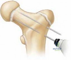
The Morrey technique utilizes an extensile lateral approach, providing unparalleled access to both the anterior and posterior compartments of the elbow while protecting critical neurovascular structures. The incision begins precisely over the lateral supracondylar ridge of the humerus, approximately 5 cm proximal to the lateral epicondyle. It is carried distally across the lateral epicondyle, ending over the subcutaneous border of the proximal ulna. Proximally, the dissection is carried directly down to the supracondylar ridge. Meticulous subperiosteal stripping is performed anterior to the anterior aspect of the humerus, elevating the brachioradialis and extensor carpi radialis longus (ECRL).
Distally, the surgeon identifies and meticulously develops the interval between the extensor carpi ulnaris (ECU) and the anconeus (the Kocher interval) to expose the lateral aspect of the elbow joint. Blunt retractors, such as Cobra or Hohmann retractors, are placed deep to the ECRL, brachioradialis, and brachialis muscles. This maneuver is absolutely critical; the brachialis muscle belly serves as a physical buffer, protecting the radial nerve and the anterior neurovascular bundle from iatrogenic injury during the anterior capsulectomy. The anconeus is reflected subperiosteally off the proximal ulna, and the distal triceps is elevated off the posterior humerus to expose the posterior compartment.
The anterior capsulectomy is performed from lateral to medial. To achieve this, the lateral collateral ligament (LCL) complex, specifically the LUCL, is often carefully reflected in a distally based flap off the lateral epicondyle. The surgeon must excise the thickened, fibrotic anterior capsule entirely. Merely incising or releasing the capsule is insufficient and significantly increases the risk of recurrent contracture. When dissecting medially, extreme caution is exercised; the dissection must remain strictly intra-articular or subperiosteal to avoid injury to the ulnar nerve and the AMCL. Following the anterior release, the posterior compartment is addressed. The olecranon fossa is cleared of all fibrotic tissue, and osteophytes at the tip of the olecranon are debrided using a high-speed burr. The final, and arguably most critical, step is the anatomical restoration of the lateral ligamentous complex. The LUCL must be securely repaired to its exact isometric origin on the lateral epicondyle using heavy nonabsorbable sutures or suture anchors to prevent postoperative posterolateral rotatory instability (PLRI).
Advanced Arthroscopic Rotator Cuff Repair and Margin Convergence
The evolution from open to arthroscopic repair has revolutionized shoulder surgery, minimizing deltoid morbidity while adhering strictly to classical biomechanical principles. The goal is to achieve a tension-free, mechanically stable footprint reconstruction that optimizes the biological environment for tendon-to-bone healing. Following a thorough diagnostic arthroscopy and meticulous footprint preparation (decorticating the greater tuberosity to a bleeding cancellous bed), the tear pattern is critically assessed. Crescent-shaped tears are highly mobile and can typically be repaired directly to the footprint using a double-row, transosseous-equivalent construct. This technique utilizes a medial row of suture anchors to compress the tendon against the articular margin, while the lateral row anchors secure the suture tails over the bursal surface, maximizing the pressurized contact area.
For massive, U-shaped or L-shaped tears that cannot be mobilized directly to the greater tuberosity without excessive tension, Burkhart introduced the revolutionary concept of "margin convergence." By utilizing side-to-side sutures to close the anterior and posterior leaves of the tear together, the surgeon effectively reduces the volume of the defect. Biomechanically, this exponential reduction in strain at the margin of the tear allows the remaining free edge of the tendon to be repaired to the bone without undue tension. Extensive capsular releases, particularly of the coracohumeral ligament and the rotator interval, are often required to mobilize these chronic, retracted tears.
Salvage Techniques: Tendon Transfers and Superior Capsule Reconstruction
When primary repair is deemed impossible due to severe tendon retraction, advanced fatty atrophy, and poor tissue quality, salvage procedures must be employed. For younger, high-demand patients with an isolated massive posterosuperior tear (supraspinatus and infraspinatus) and an intact subscapularis, the transfer of the latissimus dorsi tendon to the greater tuberosity is a highly validated option. The latissimus dorsi, an adductor and internal rotator, is transferred to act as an external rotator and a dynamic depressor of the humeral head, effectively restoring the coronal force couple.
More recently, Superior Capsule Reconstruction (SCR) utilizing dermal allograft or autologous fascia lata has emerged as a joint-preserving option. The graft is secured medially to the superior glenoid and laterally to the greater tuberosity, physically replacing the superior capsule to prevent anterosuperior escape and center the humeral head. However, in the older patient population with concomitant glenohumeral osteoarthritis (cuff tear arthropathy), the Reverse Total Shoulder Arthroplasty (RTSA) is the definitive, gold-standard treatment. By medializing the center of rotation and distalizing the humerus, the RTSA effectively recruits the deltoid muscle to elevate the arm, completely bypassing the deficient rotator cuff.
Complications, Incidence Rates, and Salvage Management
The operative management of these complex pathologies carries a significant risk profile. The surgeon must be intimately familiar with the potential complications, their incidence rates, and the appropriate salvage pathways. In the stiff elbow, neurological injury and recurrent arthrofibrosis are the most devastating complications. Iatrogenic injury to the ulnar nerve can occur during medial capsulectomy, while radial nerve palsy is a known risk of aggressive anterior retractor placement. Posterolateral rotatory instability (PLRI) occurs if the LUCL is not anatomically repaired or fails to heal, presenting with a positive pivot-shift test and profound functional limitation.
In the realm of advanced rotator cuff repair, structural failure (retear) is the most common complication, with incidence rates approaching 40-90% in massive, multi-tendon repairs. Retears are heavily influenced by patient age, tear size, and the degree of preoperative fatty infiltration. Anchor pullout, infection, and postoperative adhesive capsulitis are less frequent but equally challenging.
Table of Complications, Incidence, and Salvage Management
| Complication | Estimated Incidence | Etiology / Risk Factors | Salvage Management / Treatment |
|---|---|---|---|
| Ulnar Neuropathy (Elbow) | 5% - 10% | Traction, hematoma, or direct injury during medial capsular release. | Immediate decompression and anterior transposition if symptoms are progressive or profound. |
| Recurrent Arthrofibrosis | 10% - 20% | Inadequate initial capsulectomy, failure of postoperative CPM, non-compliance. | Revision open release vs. interposition arthroplasty; aggressive manipulation under anesthesia. |
| Iatrogenic PLRI | 2% - 5% | Failure to anatomically repair the LUCL to its isometric origin on the lateral epicondyle. | Revision ligamentous reconstruction using autograft (palmaris longus) or allograft. |
| Rotator Cuff Retear | 20% - 90% (size dependent) | Poor tendon biology, advanced age, excessive tension, smoking, non-compliance. | Asymptomatic: observe. Symptomatic: revision repair, SCR, tendon transfer, or RTSA. |
| Anterosuperior Escape | Rare (if CA arch intact) | Iatrogenic transection of the coracoacromial ligament in the setting of a massive tear. | Reverse Total Shoulder Arthroplasty (RTSA) to restore a stable fulcrum. |
| Axillary Nerve Injury | < 1% | Errant inferior portal placement or aggressive inferior capsular release. | Observation and EMG at 3 months; nerve grafting or tendon transfers if no recovery. |
The management of a failed rotator cuff repair requires a highly individualized approach. If the patient is elderly and low-demand, arthroscopic debridement and biceps tenotomy may provide sufficient pain relief without restoring strength. If the patient develops progressive cuff tear arthropathy with superior escape, RTSA is the only viable salvage option that predictably restores active elevation and alleviates pain.
Phased Post-Operative Rehabilitation Protocols
Rehabilitation Following Elbow Capsular Release
The postoperative rehabilitation following an extensive elbow release is arguably as critical as the surgical procedure itself. The protocol must aggressively combat the intense fibroproliferative response while protecting the lateral ligamentous repair.
* Immediate Phase (Days 0-3): The patient is placed in a well-padded posterior splint with the elbow in maximum extension. This position maximizes the stretch on the anterior soft tissues, which are highly prone to recurrent contracture. A perioperative indomethacin protocol (75 mg sustained release daily for 3-6 weeks) or a single dose of localized radiation (700 cGy) is administered to prevent heterotopic ossification.
* Early Motion Phase (Days 3-14): The splint is removed, and continuous passive motion (CPM) or active-assisted range of motion (AAROM) exercises are initiated. The CPM machine is utilized for 12-16 hours a day. Night splinting in maximum extension is maintained for up to 3-6 months, as the anterior capsule undergoes prolonged remodeling.
* Strengthening Phase (Weeks 6-12): Isometric and progressive resistance exercises are introduced only after the lateral ligament repair has healed sufficiently. Varus stress is strictly avoided during the early phases to protect the LUCL reconstruction.
Rehabilitation Following Advanced Rotator Cuff Repair
The rehabilitation following a massive rotator cuff repair represents a delicate balance between protecting the fragile tendon-to-bone repair and preventing postoperative adhesive capsulitis.
* Phase I: Protection Phase (Weeks 0-6): The shoulder is strictly immobilized in an abduction sling, which reduces tension on the supraspinatus repair. Only passive range of motion (PROM) is permitted, strictly avoiding any active elevation or sudden external rotation that could compromise the subscapularis or infraspinatus repairs.
* Phase II: Active-Assisted Phase (Weeks 6-10): As the initial biological healing occurs, active-assisted range of motion (AAROM) is initiated using pulleys and wand exercises. The sling is discontinued. Scapular dyskinesia is aggressively addressed by focusing on periscapular stabilizers (rhomboids, serratus anterior, trapezius).
* Phase III: Strengthening and Return to Function (Weeks 10-24): Isotonic strengthening begins with closed-chain exercises and progresses to open-chain resistance. Return to heavy manual labor or overhead sports is rarely permitted before 6 months, as the tendon remodeling phase extends well beyond a year.
Summary of Landmark Literature and Clinical Guidelines
The operative management strategies detailed in this chapter are deeply rooted in decades of rigorous biomechanical and clinical research. Mastery of this literature is essential for the academic orthopaedic surgeon.
In the realm of elbow contractures, Morrey and colleagues established the fundamental biomechanics of the elbow, defining the 100-degree functional arc of motion. Their subsequent development of the extensile lateral approach revolutionized the surgical management of the stiff elbow, proving that a single incision could safely address both anterior and posterior pathology while minimizing ulnar nerve morbidity. Hastings and Graham provided the definitive classification for heterotopic ossification about the elbow, guiding the precise timing of surgical excision based on radiographic maturation and functional limitation.
In shoulder pathology, Neer's original descriptions of subacromial impingement laid the groundwork for modern decompression techniques, while Bigliani and Morrison correlated acromial morphology directly with the incidence of rotator cuff tears. The biomechanical understanding of massive tears was fundamentally altered by Burkhart, whose concepts of the rotator cable, suspension bridge biomechanics, and margin convergence allowed surgeons to arthroscopically repair tears previously deemed inoperable. Goutallier and Fuchs established the prognostic importance of fatty infiltration, demonstrating via CT and MRI that advanced muscle degeneration is irreversible, thereby defining the absolute limits of primary tendon repair. Finally, the work of Flatow regarding the coracoacromial arch remains a critical warning to all surgeons: the preservation of the CA ligament is absolutely paramount in the setting of massive cuff deficiency to prevent the catastrophic complication of anterosuperior escape.
