Detailed Clinical Diagnosis of Lateral Epicondylitis: Patient Case & Findings
Key Takeaway
Lateral epicondylitis, or tennis elbow, is diagnosed through a detailed patient history of lateral elbow pain, often exacerbated by wrist extension. Key clinical findings include exquisite tenderness over the ECRB origin, and positive special tests like Cozen's, Mill's, and Maudsley's, which reproduce pain with resisted wrist/finger extension. Imaging typically rules out other pathology.
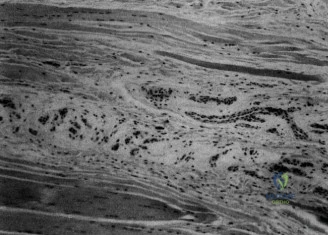
You are presented with a 48-year-old graphic designer with a 9-month history of lateral elbow pain. He has failed exhaustive conservative management, including a corticosteroid injection. Look at this MRI and describe the findings and their clinical implications for your management plan.
Candidate: The MRI shows increased signal intensity at the common extensor origin, specifically involving the ECRB tendon. There appears to be some interstitial tearing. This confirms a diagnosis of chronic lateral epicondylitis. Given the failure of non-operative treatment, I would discuss surgical intervention, such as an open debridement.
Candidates often label this simply as "tendonitis" or "inflammation." They fail to identify the specific anatomical involvement (ECRB) versus the EDC, or they neglect to comment on the integrity of the Lateral Ulnar Collateral Ligament (LUCL). Failing to mention that the pathology is "angiofibroblastic tendinosis" rather than inflammatory is a significant oversight at the FRCS level.
The MRI demonstrates classic features of chronic angiofibroblastic tendinosis of the ECRB origin. Specifically, there is T2-hyperintensity representing mucoid degeneration and a high-grade partial-thickness interstitial tear. I would highlight that the LUCL is intact, which is critical to note before planning surgery. I would categorize this as a recalcitrant case, and given the 9-month duration and failure of formal eccentric loading/physiotherapy, I would recommend surgical debridement (Nirschl procedure) to remove the pathological tissue and stimulate a healing response via decortication of the epicondyle.
During your preoperative planning, how would you distinguish between lateral epicondylitis and Radial Tunnel Syndrome (RTS) clinically?
Candidate: Lateral epicondylitis presents with pain at the ECRB origin, while RTS is caused by nerve compression. In RTS, the patient usually has pain further down the forearm, and resisted supination reproduces their symptoms.
Missing the exact anatomical localization. Borderline candidates fail to specify the "Mobile Wad of Henry" and don't clearly distinguish the difference between testing the ECRB (resisted wrist/middle finger extension) versus testing the supinator/PIN (resisted supination).
I distinguish them based on the site of maximal tenderness and provocative testing. 1. Localization: Epicondylitis is localized to the ECRB origin (1-2cm distal to the lateral epicondyle), whereas RTS tenderness is 3-4cm distal, over the radial tunnel/supinator muscle. 2. Provocation: Epicondylitis is reproduced by resisted wrist extension and Maudsley's test (resisted middle finger extension). RTS is reproduced by resisted forearm supination with the elbow extended, which compresses the PIN at the Arcade of Frohse. 3. Clinical note: I would emphasize that RTS and epicondylitis can coexist, and failing to diagnose RTS is a common cause of "failed" lateral epicondylitis surgery.
You have decided to proceed with an open surgical debridement. What are the specific intraoperative anatomical risks, and how do you mitigate them?
Candidate: You must avoid cutting the LUCL, as that causes instability. You also have to be careful with the radial nerve and the posterior interosseous nerve.
The candidate mentions general structures without defining the "safe zone" or specific landmarks. They often forget the cutaneous branches (Posterior Antebrachial Cutaneous Nerve) which can lead to postoperative neuroma and patient dissatisfaction.
There are three primary risks: 1. LUCL Injury: I avoid this by staying anterior to the equator of the radiocapitellar joint. The LUCL originates posterior and distal to the ECRB footprint; violating it leads to posterolateral rotatory instability. 2. Posterior Antebrachial Cutaneous Nerve (PABCN): This branch is vulnerable during the superficial skin incision and dissection. I identify and protect it using blunt retraction to prevent painful postoperative neuromas. 3. PIN Injury: While the PIN is deep to the supinator, I avoid overly deep or medial dissection once I have reached the epicondylar bone to ensure no injury to the nerve as it enters the arcade of Frohse.
