Elbow Collateral Ligament Instability: Epidemiology, Anatomy, and Biomechanics
Key Takeaway
Elbow collateral ligament instability involves injuries to the medial (UCL) and lateral (LCL) complexes, critical for elbow function. UCL injuries often affect overhead athletes due to valgus stress, while LCL injuries typically cause posterolateral rotatory instability from trauma like FOOSH. Understanding their distinct anatomy and biomechanics is paramount for diagnosis and treatment.
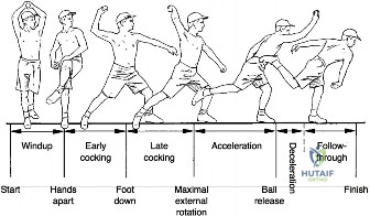
A 24-year-old professional baseball pitcher presents with a 4-month history of medial-sided elbow pain and a noticeable decline in pitching velocity. He reports a "dead arm" sensation during the late cocking phase. Clinical examination reveals tenderness over the medial epicondyle and a positive valgus stress test at 30 degrees of flexion. Please interpret this clinical picture and discuss your further management.
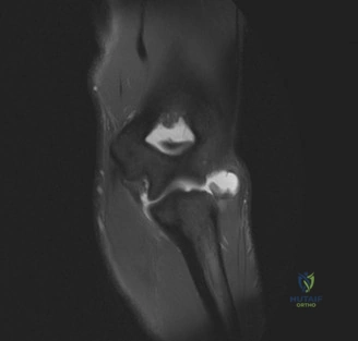
Candidate: The presentation is classic for a Medial Collateral Ligament (UCL) injury, likely an anterior bundle partial tear or attenuation due to repetitive microtrauma. I would order MRI to confirm the pathology and assess for associated injuries. Given his profession, if conservative management fails, I would discuss UCL reconstruction (e.g., modified docking technique) using an autograft like the palmaris longus.
Candidates often fail to quantify the clinical test or mention specific imaging. Simply saying "MRI" is insufficient; you must discuss the role of stress radiographs to confirm instability. Failing to mention the ulnar nerve—its proximity and potential for symptoms—is a major oversight in any medial elbow scenario.
Start with a structured approach: History (overhead athlete), Physical Exam (valgus laxity, ulnar nerve status), and Imaging (Stress X-rays to compare opening with the contralateral side; MRI to confirm the tear and look for 'T-sign' or signal changes at the sublime tubercle). Mention that for a high-demand athlete, if he fails 3-6 months of conservative management (kinetic chain optimization), UCL reconstruction is indicated. Note the ulnar nerve: it must be identified and protected, with transposition only if indicated by preoperative symptoms or instability.
Following a fall onto an outstretched hand (FOOSH), this patient developed chronic elbow instability. Clinical exam shows a positive pivot shift test. You are planning an anatomical LUCL reconstruction. Explain the surgical anatomy and the key technical objective to avoid iatrogenic complications.
Candidate: The LUCL is the primary stabilizer against posterolateral rotatory instability (PLRI). The key technical objective is identifying the isometric point on the lateral epicondyle. During dissection, the posterior interosseous nerve (PIN) is the main structure at risk. It lies anterior to the radial head and pierces the supinator 3-4cm distal to the joint; I would remain posterior to the radial head to protect it.
Candidates often confuse the LUCL with the radial collateral ligament (RCL). They also frequently omit the importance of limb position during tensioning—tensioning in full supination rather than pronation leads to an overtightened construct and loss of rotation.
Define the anatomy: The LUCL originates from the lateral epicondyle and inserts on the supinator crest of the ulna. Crucially, emphasize that reconstruction must be tensioned with the elbow in 60-90 degrees of flexion and the forearm in pronation to restore stability while maintaining rotational arc. Mention the "safe zone" for the PIN and note that the common extensor origin should be elevated or split carefully to preserve the repair integrity.
