Elbow Calcific Tendonitis: Comprehensive Diagnosis, Clinical Presentation & Phases
Key Takeaway
Elbow calcific tendonitis is diagnosed by evaluating severe, localized pain, often disproportionate to trauma, with activity or at rest. Key findings include pinpoint tenderness over specific elbow tendons (e.g., triceps, epicondyles) during clinical examination. Understanding the distinct phases, especially the acute resorptive phase, is crucial, as it typically correlates with the most intense symptoms and guides diagnostic imaging.
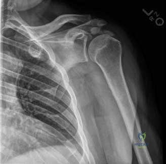
A 45-year-old patient presents to your clinic complaining of a 3-week history of severe, unrelenting pain in their right elbow. They report no history of trauma. The pain is worst at night and is not responding to oral NSAIDs. On examination, they have localized, exquisite tenderness at the tip of the olecranon. You order the following radiograph.
What is your primary differential diagnosis, and how would you describe the imaging findings?
Candidate: The primary diagnosis is calcific tendonitis of the triceps. The X-ray shows a radiodensity at the insertion of the triceps tendon. It could also be olecranon bursitis or an avulsion fracture.
Failing to mention the "phase" of the condition or ignoring the clinical urgency. A poor candidate treats this like a standard tendonopathy and fails to identify the resorptive phase implications, or forgets to differentiate it from septic olecranon bursitis—a critical clinical "must-not-miss."
The diagnosis is calcific tendonitis of the triceps insertion. The radiograph shows an amorphous, radiopaque deposit adjacent to the olecranon. Given the clinical presentation of acute, severe, constant pain, the patient is likely in the resorptive phase of the disease. I must differentiate this from septic olecranon bursitis by assessing for systemic symptoms, fluctuance, and potentially inflammatory markers if the diagnosis remains in doubt.
The patient is significantly distressed by the pain. They have failed two weeks of oral ibuprofen. What is your next management step, and what specific procedure would you discuss with them?
Candidate: I would offer them a corticosteroid injection. If that fails, I would suggest surgery to remove the calcium.
Suggesting a "blind" corticosteroid injection. This is a significant error; intra-tendinous steroid injection carries a high risk of tendon weakening or rupture. Furthermore, skipping the mention of "barbotage" misses the most effective evidence-based intervention for this phase.
I would propose ultrasound-guided barbotage (lavage and aspiration). This is the gold standard for symptomatic relief in the resorptive phase, as it mechanically removes the inflammatory calcium crystals. I would emphasize to the patient that this is minimally invasive. I would avoid direct intra-tendinous corticosteroid injection due to the risk of tendon rupture; however, a peri-tendinous injection can be performed as an adjunct for pain control.
At what point would you consider open surgical excision, and what specific intra-operative precautions are necessary?
Candidate: I would consider surgery if they have had pain for over 6 months despite conservative care. Intra-operatively, I would make an incision, find the calcium, and take it out.
Failing to mention the proximity of the ulnar nerve (for medial or posterior approaches) or the importance of intra-operative imaging. Simply saying "take it out" is not enough—you must mention confirming complete excision to prevent recurrence.
Surgery is reserved for cases failing 6-12 months of comprehensive non-operative management. Precautions include: 1) Meticulous identification and protection of neurovascular structures (specifically the ulnar nerve if approaching the posterior or medial elbow). 2) Use of intra-operative fluoroscopy or ultrasound to ensure complete excision of the deposit, as residual calcium is a common cause of persistent symptoms. 3) Careful handling of the tendon to avoid creating a large defect requiring formal repair.
