Surgical Management of Biceps Brachii Tendon Displacement: A Comprehensive Operative Guide
Key Takeaway
Biceps brachii tendon displacement frequently presents alongside rotator cuff pathology. Surgical management depends on the integrity of the subscapularis and the transverse humeral ligament. While isolated transverse humeral ligament repair is historically described, modern evidence strongly favors biceps tenodesis. This guide details the anterosuperior and deltopectoral approaches, arthroscopic-assisted subpectoral tenodesis, and postoperative rehabilitation protocols to optimize functional outcomes in patients with long head of the biceps tendon instability.
Comprehensive Introduction and Patho-Epidemiology
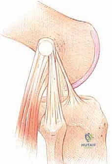
Displacement, subluxation, or frank dislocation of the long head of the biceps (LHB) tendon is a highly complex pathological entity that represents a significant source of anterior shoulder morbidity. This condition rarely occurs in an isolated anatomical vacuum; rather, it is intimately tied to the structural integrity of the surrounding rotator cuff and the capsuloligamentous structures of the anterior shoulder. The stability of the LHB within the bicipital groove (intertubercular sulcus) is dynamically and statically maintained by a sophisticated anatomical construct known as the "biceps pulley." This pulley system is composed of the superior glenohumeral ligament (SGHL), the coracohumeral ligament (CHL), and the superior fibers of the subscapularis tendon. Historically, the transverse humeral ligament (THL) was thought to be the primary restraint; however, modern anatomical studies have definitively proven that the THL is merely a fascial continuation of the subscapularis and supraspinatus, acting only as a secondary, relatively weak restraint over the intertubercular groove.
The epidemiology of LHB displacement demonstrates a strong correlation with advancing age, chronic overhead activity, and degenerative rotator cuff disease. True isolated displacement of the biceps tendon without concomitant rotator cuff or pulley pathology is exceedingly rare, accounting for less than 2% of all biceps pathologies. The vast majority of these lesions are identified in patients over the age of 50 who present with concomitant tears of the subscapularis or supraspinatus tendons. In younger, athletic populations, LHB displacement is typically traumatic in origin, often resulting from a forceful eccentric contraction of the biceps during a fall or an extreme external rotation and abduction injury that simultaneously avulses the subscapularis insertion and the medial pulley sling.
When the medial stabilizing structures of the biceps pulley are disrupted, the LHB tendon inevitably subluxates or dislocates medially over the lesser tuberosity. This displacement alters the biomechanical vectors of the glenohumeral joint. A medially dislocated biceps tendon acts as an abrasive bowstring across the anterior joint, leading to persistent anterior shoulder pain, mechanical clicking, and progressive attrition of the subscapularis tendon due to chronic friction. This phenomenon is often referred to as a "hidden lesion" because the dislocated tendon can obscure underlying subscapularis tears during standard arthroscopic evaluation from a posterior portal unless a systematic and thorough examination of the anterior compartment is performed.
The evolution of surgical management for LHB displacement reflects a paradigm shift in orthopedic understanding of shoulder biomechanics. Historically, surgical treatment involved attempting to relocate the tendon back into the bicipital groove and primarily repairing the transverse humeral ligament or constructing a soft-tissue sling. However, long-term outcome studies demonstrated unacceptably high failure rates, persistent anterior shoulder pain, and recurrent dislocations with these joint-preserving techniques. Consequently, the contemporary orthopedic consensus recognizes that addressing the underlying pulley lesion via primary repair without addressing the diseased LHB tendon is futile. Today, tenodesis or tenotomy of the LHB, coupled with appropriate rotator cuff repair, has become the unequivocal gold standard for managing LHB displacement.
Detailed Surgical Anatomy and Biomechanics
A profound mastery of the surgical anatomy of the anterior shoulder is an absolute prerequisite for the operative management of LHB displacement. The long head of the biceps tendon originates intra-articularly from the supraglenoid tubercle and the superior glenoid labrum. It traverses the glenohumeral joint obliquely, passing over the humeral head before exiting the joint capsule to enter the bicipital groove. The bicipital groove itself exhibits significant morphological variability among individuals; a shallow groove or a medial wall angle of less than 30 degrees substantially predisposes the patient to tendon subluxation. Within the groove, the tendon is enveloped by a synovial sheath that communicates directly with the glenohumeral joint, explaining why intra-articular inflammatory processes, such as adhesive capsulitis or rheumatoid arthritis, frequently manifest with severe bicipital tenosynovitis.
The architecture of the biceps pulley is an intricate confluence of ligamentous and tendinous structures. The coracohumeral ligament (CHL) originates from the base of the coracoid process and bifurcates into two distinct bands: the medial band inserts onto the lesser tuberosity, blending with the subscapularis, while the lateral band inserts onto the greater tuberosity, blending with the supraspinatus. The superior glenohumeral ligament (SGHL) runs an oblique course from the upper labrum to the lesser tuberosity, forming a U-shaped suspension sling around the LHB tendon alongside the CHL. The superior-most fibers of the subscapularis tendon pass beneath this sling, providing a critical dynamic buttress against medial tendon displacement. Disruption of this complex, particularly the medial sling (SGHL and medial CHL), removes the primary barrier to medial subluxation.
The vascularity and innervation of this region dictate the safe zones for surgical dissection. The primary blood supply to the proximal biceps tendon and the surrounding pulley structures is derived from the ascending branch of the anterior circumflex humeral artery (often referred to as the arcuate artery of Laing). This vessel courses parallel to the lateral aspect of the bicipital groove. Iatrogenic injury to this artery during deep dissection, particularly during subpectoral approaches, can result in profound, difficult-to-control hemorrhage and subsequent hematoma formation. Neurologically, the musculocutaneous nerve is the most critical structure at risk during open subpectoral tenodesis. It branches from the lateral cord of the brachial plexus and typically enters the coracobrachialis muscle 3 to 5 centimeters distal to the tip of the coracoid process, though anatomical variants exist where the nerve enters much more proximally.
Biomechanically, the exact role of the LHB in glenohumeral kinematics remains a subject of academic debate. While it functions as a weak flexor of the shoulder and a supinator of the forearm, its primary role at the shoulder joint is believed to be a dynamic depressor of the humeral head, particularly in the setting of massive rotator cuff tears. However, when the LHB displaces medially, its vector of force is drastically altered. Instead of depressing the humeral head, the subluxated tendon generates an anteriorly directed shear force, acting as a "bowstring" that mechanically degrades the subscapularis footprint. This altered kinematic state not only exacerbates anterior shoulder instability but also generates severe nociceptive feedback due to the rich sensory innervation of the bicipital sheath and the surrounding synovium.
Exhaustive Indications and Contraindications
The decision to proceed with surgical intervention for LHB displacement must be predicated on a meticulous correlation of patient symptomatology, functional demands, and advanced imaging findings. Surgical management is primarily indicated for patients who present with refractory anterior shoulder pain, mechanical symptoms (such as painful clicking or snapping with internal and external rotation), and objective evidence of LHB subluxation or dislocation. Non-operative management—comprising targeted physical therapy, nonsteroidal anti-inflammatory drugs (NSAIDs), and judicious ultrasound-guided corticosteroid injections into the bicipital sheath—should typically be exhausted over a 3- to 6-month period before considering surgical intervention, unless a concomitant acute, repairable rotator cuff tear is identified.
The choice between biceps tenodesis and biceps tenotomy represents a critical branch point in the surgical algorithm. Biceps tenodesis is the procedure of choice for younger, highly active patients, manual laborers, and athletes. Tenodesis restores the physiological resting length-tension relationship of the biceps muscle, thereby preserving maximal supination and elbow flexion strength. Furthermore, it prevents the distal migration of the muscle belly, avoiding the cosmetically unappealing "Popeye" deformity and mitigating the risk of subjective muscle cramping, which is reported in up to 20% of tenotomy patients. Conversely, simple arthroscopic biceps tenotomy is an elegantly simple, highly effective option for low-demand, elderly patients, or those with significant medical comorbidities where minimizing operative time is paramount.
Contraindications to surgical intervention, and specifically to biceps tenodesis, must be carefully weighed. Absolute contraindications include active local or systemic infection, severe medical comorbidities precluding safe anesthesia, and the presence of a complex regional pain syndrome (CRPS) affecting the operative extremity. Relative contraindications include profound glenohumeral stiffness (adhesive capsulitis); in such cases, the stiffness should ideally be addressed via physical therapy or arthroscopic capsular release prior to or concurrent with the LHB intervention. Additionally, severe osteoporosis may be a relative contraindication to certain types of interference screw fixation due to the risk of hardware pull-out or iatrogenic humeral shaft fracture, necessitating the use of alternative fixation methods such as cortical buttons or suture anchors.
| Parameter | Biceps Tenodesis | Biceps Tenotomy | Non-Operative Management |
|---|---|---|---|
| Primary Indications | Active patients (<65 yrs), laborers, athletes, cosmetic concern | Elderly, low-demand patients (>65 yrs), massive irreparable cuff tears | First-line for isolated tendinosis/mild subluxation without cuff tear |
| Advantages | Preserves strength, prevents Popeye deformity, avoids cramping | Technically simple, rapid recovery, no implant-related complications | Avoids surgical risks, preserves native anatomy |
| Disadvantages | Longer operative time, implant costs, risk of humerus fracture | Cosmetic deformity, potential loss of supination strength, cramping | High failure rate for frank dislocations, progressive cuff attrition |
| Contraindications | Severe osteoporosis (for screw fixation), active infection | High-demand athletes requiring peak supination torque | Acute, repairable subscapularis avulsions with subluxed LHB |
Pre-Operative Planning, Templating, and Patient Positioning
Thorough pre-operative planning begins with a comprehensive clinical examination designed to isolate LHB pathology from concomitant shoulder disorders. Provocative testing is essential. Speed’s test and Yergason’s test are classic maneuvers for assessing bicipital groove pathology, though their sensitivity and specificity are variable. More importantly, the surgeon must systematically evaluate the subscapularis tendon, given its intimate relationship with the medial biceps pulley. The Bear Hug test, Belly Press test, and Lift-Off test are mandatory. A positive result on these tests in the presence of anterior shoulder pain is highly suspicious for a combined subscapularis tear and LHB subluxation, a combination that dictates an operative approach capable of addressing both lesions simultaneously.
Advanced imaging is the cornerstone of pre-operative templating. While standard orthogonal radiographs (true anteroposterior, scapular Y, and axillary lateral views) are necessary to rule out osseous pathology, a specialized Fisk view or a bicipital groove view can be obtained to assess groove depth and the presence of osteophytes. However, Magnetic Resonance Imaging (MRI), preferably an MR arthrogram, is the gold standard. Axial T2-weighted sequences are meticulously scrutinized at the level of the lesser tuberosity to evaluate the position of the LHB. An "empty groove" sign, combined with a medially displaced tendon resting over the subscapularis footprint, is pathognomonic for LHB dislocation. Furthermore, the surgeon must template the anticipated size of the tendon to ensure the availability of appropriately sized interference screws and reamers (typically ranging from 7 to 9 millimeters).
Anesthetic management significantly influences patient positioning and operative efficiency. A multimodal approach utilizing an ultrasound-guided interscalene brachial plexus block combined with general anesthesia is preferred. This provides profound intra-operative muscle relaxation, crucial for manipulating the arm during tenodesis, and ensures excellent immediate post-operative analgesia. The anesthesiologist must be cautioned regarding the potential for phrenic nerve palsy associated with interscalene blocks, and alternative regional techniques (such as a supraclavicular block) may be considered in patients with severe pre-existing pulmonary compromise.
Patient positioning is dictated by the surgeon's preference and the planned surgical approach. The modified beach-chair position is the most versatile, allowing seamless transition from arthroscopic evaluation to an open deltopectoral or subpectoral approach. The patient is placed in approximately 45 to 60 degrees of upright tilt, with the operative arm completely free to allow full range of motion. The head must be rigidly secured in a neutral position to prevent cervical spine hyperextension or lateral flexion, which could stretch the contralateral brachial plexus. Alternatively, the lateral decubitus position can be utilized, particularly if the surgeon prefers this setup for concomitant arthroscopic rotator cuff repair; however, converting to an open subpectoral tenodesis from the lateral decubitus position is ergonomically challenging and often requires repositioning.
Step-by-Step Surgical Approach and Fixation Technique
Arthroscopic Evaluation and Intra-Articular Release
Regardless of the planned open approach for tenodesis, the procedure universally begins with a comprehensive diagnostic arthroscopy. Standard posterior and anterior portals are established. The arthroscope is introduced posteriorly, and a systematic 15-point diagnostic tour of the glenohumeral joint is performed. The biceps pulley is meticulously probed. The "comma sign"—a distinct comma-shaped arc of tissue representing the avulsed superior glenohumeral ligament and coracohumeral ligament complex—is pathognomonic for a combined subscapularis tear and LHB subluxation. The LHB tendon is drawn into the joint using a probe to assess for hidden tendinosis, partial tearing, or hourglass deformity.
Once the pathology is confirmed, an arthroscopic intra-articular tenotomy is performed. Using an electrothermal ablation device or arthroscopic scissors introduced through the anterior portal, the LHB is transected as close to its origin on the superior labrum as possible. Care must be taken not to destabilize the superior labrum anterior to posterior (SLAP) complex during this release. If the tendon is severely hypertrophied (an "hourglass" biceps), it may become incarcerated within the joint and refuse to retract into the groove. In such cases, the tendon must be aggressively pulled distally during the open phase or excised piecemeal arthroscopically to prevent a painful intra-articular stump.
Open Subpectoral Tenodesis
The open subpectoral approach is widely considered the gold standard for LHB tenodesis, as it entirely removes the tendon from the bicipital groove, eliminating any potential for residual groove-related pain. The patient’s arm is abducted and externally rotated. A 2 to 3 centimeter longitudinal incision is made centered over the inferior border of the pectoralis major tendon, hidden within the axillary fold for optimal cosmesis. Subcutaneous dissection is carried down to the pectoralis major fascia. The inferior border of the pectoralis major is identified, and a blunt retractor (such as a Chandler or right-angle retractor) is placed under the muscle belly, retracting it superiorly. The conjoint tendon is identified medially and gently retracted medialward, taking extreme care to avoid traction neuropraxia to the musculocutaneous nerve.
Deep to the pectoralis major, the bicipital sheath is identified. The sheath is incised longitudinally, and the LHB tendon is delivered into the wound using a right-angle clamp. The diseased proximal portion of the tendon is excised. The remaining healthy tendon is then secured. A high-strength, non-absorbable suture (e.g., #2 FiberWire) is used to place a locking whipstitch (such as a Krackow or grasping stitch) over the distal 2.5 centimeters of the tendon. The humerus is exposed at the base of the bicipital groove, approximately 1 centimeter distal to the inferior border of the pectoralis major. A guide pin is placed perpendicularly into the center of the humeral shaft, and a reamer (sized to match the diameter of the prepared tendon, typically 7 or 8 mm) is used to create a unicortical bone socket.
Suprapectoral and Deltopectoral Approaches
In scenarios where a massive rotator cuff tear, particularly a large subscapularis avulsion, requires an open or mini-open repair, a suprapectoral or standard deltopectoral approach may be utilized. The deltopectoral interval is developed between the deltoid and the pectoralis major, retracting the cephalic vein laterally. The transverse humeral ligament is incised to expose the subluxated tendon. In the suprapectoral technique, the tenodesis is performed within the proximal aspect of the bicipital groove. While this allows for simultaneous repair of the subscapularis tendon through the same window, it carries a higher risk of persistent groove pain if the tendon is not adequately tensioned or if the groove itself is stenotic or osteophytic.
If an anterosuperior deltoid-splitting approach is utilized (often for concomitant supraspinatus pathology), the split must not extend beyond 5 centimeters distal to the lateral edge of the acromion. Extending this split places the axillary nerve at imminent risk of transection, a catastrophic complication resulting in profound deltoid paralysis. A heavy stay suture is placed at the distal apex of the split to prevent inadvertent propagation during retraction. The tendon is identified, tenotomized proximally, and tenodesed into the proximal groove using suture anchors, followed by the requisite rotator cuff repair.
Fixation Biomechanics and Techniques
The biomechanical integrity of the tenodesis construct is paramount for early rehabilitation and optimal functional outcomes. Interference screw fixation (subpectoral) provides the highest initial pull-out strength and stiffness compared to suture anchors. The prepared tendon is introduced into the unicortical socket using a specialized driver. A bio-composite or PEEK interference screw, typically the same diameter as the reamer or 0.5 mm larger, is advanced over the tendon into the socket. The surgeon must maintain strict attention to the length-tension relationship; the superior border of the biceps muscle belly should rest at the level of the inferior border of the pectoralis major tendon when the elbow is fully extended.
Alternatively, a cortical button technique (tension-slide technique) can be employed. This involves drilling a small bicortical tunnel through the humerus. The button is passed through the tunnel and flipped on the posterior cortex. The sutures are then tensioned, drawing the tendon into the unicortical anterior socket. This technique relies on cortical fixation rather than cancellous interference and is particularly advantageous in patients with osteoporotic bone where an interference screw might strip or fail to achieve adequate purchase. Suture anchors remain a viable option, particularly for suprapectoral tenodesis, but require a meticulously tied knot construct and generally exhibit lower ultimate load-to-failure rates in biomechanical testing.
Complications, Incidence Rates, and Salvage Management
While biceps tenodesis is a highly successful procedure with patient satisfaction rates frequently exceeding 90%, the surgeon must be acutely aware of potential complications and possess the technical acumen to manage them. Complications can be broadly categorized into cosmetic, mechanical, neurologic, and infectious etiologies. A thorough understanding of these risks is essential for informed pre-operative patient counseling and vigilant post-operative monitoring.
The most common complication following biceps surgery is the cosmetic "Popeye" deformity, which results from the distal migration of the biceps muscle belly. In the setting of an isolated tenotomy, the incidence of a Popeye deformity ranges from 15% to 45%, depending on the patient's body mass index and muscle tone. Following a tenodesis, a Popeye deformity indicates a failure of the fixation construct or inadequate intra-operative tensioning. If a tenodesis fails early in the post-operative period (pull-out), salvage management typically involves a revision open subpectoral tenodesis, often requiring a larger interference screw, a cortical button construct, or securing the tendon directly to the pectoralis major fascia if the tendon length is insufficient to reach the humerus.
Neurologic injury is a rare but devastating complication. The musculocutaneous nerve is at the highest risk during the open subpectoral approach. This neuropraxia is almost universally caused by aggressive, prolonged medial retraction of the conjoint tendon rather than direct transection. The incidence is reported to be less than 1%, but it manifests as severe weakness in elbow flexion and sensory deficits over the lateral forearm. Most cases are transient neuropraxias that resolve with observation over 3 to 6 months. If an anterosuperior approach is used, the axillary nerve is at risk if the deltoid split exceeds 5 cm. Vascular complications, specifically hematoma formation from the ascending branch of the anterior circumflex humeral artery, occur in 1-2% of cases and may require operative evacuation if they cause compressive symptoms or impending skin necrosis.
| Complication | Estimated Incidence | Etiology / Risk Factor | Salvage / Management Strategy |
|---|---|---|---|
| "Popeye" Deformity | 15-45% (Tenotomy) 1-3% (Tenodesis) |
Fixation failure, inadequate tensioning, simple tenotomy | Revision open tenodesis, soft-tissue tethering to pec major |
| Persistent Groove Pain | 5-10% (Suprapectoral) | Tendon retained in stenotic groove, inadequate resection | Revision to subpectoral tenodesis, arthroscopic groove decompression |
| Musculocutaneous Neuropraxia | < 1% | Aggressive medial retraction of conjoint tendon | Observation, EMG at 6 weeks; usually resolves spontaneously |
| Humeral Shaft Fracture | < 0.5% | Oversized reamer, eccentric drilling, severe osteoporosis | Open reduction internal fixation (ORIF) with long locking plate |
| Adhesive Capsulitis | 2-5% | Prolonged immobilization, poor pain control | Aggressive physical therapy, intra-articular steroids, arthroscopic release |
Phased Post-Operative Rehabilitation Protocols
The post-operative rehabilitation protocol following surgical management of LHB displacement must be meticulously structured, phased, and tailored to the specific surgical interventions performed. Crucially, if a concomitant rotator cuff repair or subscapularis repair was executed alongside the biceps tenodesis, the rehabilitation timeline is universally dictated by the healing constraints of the rotator cuff, which requires a more conservative progression than an isolated tenodesis. The primary philosophy of rehabilitation is to balance the protection of the healing tenodesis construct with the prevention of glenohumeral adhesive capsulitis.
Phase I: Protection and Early Passive Motion (Weeks 0–4)
The immediate post-operative phase focuses on protecting the surgical repair and managing inflammation. The patient is placed in a standardized shoulder immobilizer or sling, which is worn continuously, including during sleep, for the first 4 weeks. Passive range of motion (PROM) of the glenohumeral joint is initiated within the first week to prevent capsular contracture. This includes passive forward elevation, external rotation (limited to 30 degrees if a subscapularis repair was performed), and internal rotation. Active elbow flexion and active forearm supination are strictly prohibited, as these motions directly load the biceps tenodesis site. Passive elbow flexion and extension are permitted and encouraged to prevent elbow stiffness.
Phase II: Active-Assisted and Active Motion (Weeks 4–8)
At the 4-week mark, the sling is gradually weaned and discontinued. The rehabilitation focus shifts toward restoring active kinematics. Active-assisted range of motion (AAROM) exercises, such as pulley systems and wand exercises, are introduced and progressively transitioned to full active range of motion (AROM) of the shoulder. Regarding the biceps, gentle, unresisted active elbow flexion and active supination are initiated. The patient is instructed to perform these movements in a controlled manner, avoiding any sudden, jerky motions. Submaximal isometric exercises for the periscapular stabilizers and the intact rotator cuff musculature are also incorporated to re-establish neuromuscular control.
Phase III: Progressive Strengthening (Weeks 8–12)
Once full, painless active range of motion is achieved, the strengthening phase begins. Isotonic strengthening of the rotator cuff, deltoid, and periscapular muscles is advanced using resistance bands and light free weights. Progressive resisted exercises specifically targeting the biceps brachii are initiated at the 8-week mark. This begins with light resistance (e.g., 1- to 2-pound dumbbells) for elbow flexion and supination, focusing on high repetitions to build muscular endurance rather than peak strength. The physical therapist must closely monitor the patient for any signs of pain at the bicipital groove or the subpectoral tenodesis site, which would necessitate a temporary reduction in resistance.
Phase IV: Advanced Strengthening and Return to Activity (Months 3–6)
The final phase of rehabilitation bridges the gap between basic functional recovery and a return to high-demand activities. Advanced strengthening, including plyometrics and eccentric loading of the biceps and rotator cuff, is introduced. For athletes, sport-specific training (such as throwing programs or overhead racket mechanics) is initiated. Clearance for a return to heavy manual labor or competitive overhead sports is typically granted between 4 and 6 months post-operatively. The criteria for unrestricted clearance include full, symmetric, and painless range of motion, normal scapulothoracic rhythm, and isokinetic strength testing demonstrating at least 90% strength compared to the contralateral, uninjured extremity.
Summary of Landmark Literature and Clinical Guidelines
The contemporary surgical management of long head of the biceps displacement is heavily informed by decades of rigorous anatomical, biomechanical, and clinical research. A foundational understanding of this literature is essential for the practicing orthopedic surgeon to justify clinical decision-making and adhere to evidence-based best practices. Historically, the treatment of LHB subluxation was controversial, but landmark studies have forged the current consensus.
The anatomical definition of the biceps pulley and its pathological variations were definitively categorized by Habermeyer and Walch in the late 1990s and early 2000s. Walch's classification of pulley lesions highlighted the intrinsic relationship between the superior glenohumeral ligament, the coracohumeral ligament, and the subscapularis tendon. Their work established the "hidden lesion" concept, proving that medial subluxation of the LHB is almost universally accompanied by a partial or complete articular-sided subscapularis tear, fundamentally shifting the surgical approach from isolated THL repair to comprehensive anterior compartment reconstruction.
The debate between biceps tenodesis and tenotomy was significantly clarified by the work of Boileau and Werner. Boileau's extensive clinical series demonstrated that while tenotomy provides excellent pain relief, it carries a high risk of cosmetic deformity and fatigue cramping in active patients. Werner’s biomechanical studies further elucidated that tenodesis preserves the physiological length-tension curve of the biceps, maintaining peak supination torque, which is critically important for manual laborers. Consequently, current American Academy of Orthopaedic Surgeons (AAOS) and American Shoulder and Elbow Surgeons (ASES) guidelines strongly recommend tenodesis over tenotomy for patients under the age of 65 or those engaged in heavy occupational or athletic demands.
Biomechanical optimization of tenodesis fixation has been extensively researched by Mazzocca and colleagues. Their landmark cadaveric studies compared various fixation constructs, including interference screws, cortical buttons, and suture anchors. They conclusively demonstrated that subpectoral interference screw fixation provides superior ultimate load-to-failure and minimal cyclic displacement compared to other techniques, establishing it as the biomechanical gold standard. However, subsequent clinical outcome studies have shown that while biomechanical differences exist, clinical success rates between subpectoral interference screws and cortical button techniques are largely equivalent, allowing surgeons to tailor the fixation method to the patient's specific bone quality and anatomical constraints.
