Unraveling Infraspinatus and Teres Rotator Cuff Tears
Key Takeaway
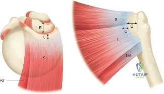
Your ultimate guide to Unraveling Infraspinatus and Teres Rotator Cuff Tears starts here. The rotator cable is a thick band of tissue, a thickening of the coracohumeral ligament. It runs perpendicular to muscle fibers near the supraspinatus and infraspinatus, extending to the inferior edge of the infraspinatus, adjacent to the teres minor. Its proposed function is to distribute forces on the humeral head, acting like a suspension bridge for the rotator cuff.
A 58-year-old active manual laborer presents with a 4-month history of progressive right shoulder weakness and pain. He specifically complains of an inability to open heavy doors and difficulty with overhead tasks. On examination, he demonstrates a positive Hornblower’s sign. What is your clinical suspicion, and what specific anatomical structure is primarily implicated?
Candidate: I suspect a massive posterosuperior rotator cuff tear involving the infraspinatus and teres minor. The positive Hornblower's sign specifically points to severe teres minor deficiency, as the patient is unable to maintain external rotation in 90 degrees of abduction.
Candidates often confuse the "External Rotation Lag Sign" with "Hornblower's Sign." They may simply state "rotator cuff tear" without differentiating the specific tendons involved, or fail to mention the loss of posterior dynamic stability, which is the functional consequence of teres minor involvement.
The candidate should identify a massive posterosuperior cuff tear. They must explain: 1) Hornblower's sign is pathognomonic for teres minor (and infraspinatus) deficiency. 2) The patient has lost the ability to generate external rotation torque in the abducted position. 3) Crucially, the teres minor is the "sister muscle" to the deltoid, and its loss predisposes the joint to posterior-inferior subluxation due to the loss of the posterior force couple.
You are planning an arthroscopic repair for a chronic, massive posterosuperior tear. You are evaluating the patient's MRI. What are the key prognostic indicators you look for, and how do they influence your decision-making?
Candidate: I assess the Goutallier grade for fatty infiltration and the degree of muscle atrophy. If the grade is 3 or 4, the repair is likely to fail or have a high re-tear rate, potentially making the tear irreparable. I also look at retraction (Patte classification).
Failing to mention that these changes are often irreversible. Candidates often stop at "the repair is difficult" without explaining that for Goutallier 3-4, they should pivot to discussing alternative procedures like SCR or reverse total shoulder arthroplasty (rTSA).
A structured answer: 1) Fatty Infiltration (Goutallier): Grades 3-4 indicate high failure rates for direct repair. 2) Retraction (Patte): Stage 3 (retraction to the glenoid) suggests significant loss of tension-free excursion. 3) Clinical correlation: If irreparable (high Goutallier, severe atrophy), the discussion shifts to salvage options like Reverse TSA or Superior Capsule Reconstruction (SCR) rather than attempting a primary repair that is prone to failure.
During the arthroscopic repair of this infraspinatus and teres minor tear, you decide to use the lateral decubitus position. What are the specific intraoperative considerations for patient safety and joint access in this position?
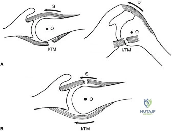
Candidate: In lateral decubitus, you get good distraction of the joint, but you must be careful with traction weight—usually 10-15 lbs—to avoid brachial plexus neurapraxia. You also need to watch for pressure points on the non-operative side.
Neglecting the "Posterior Working Portals." Candidates often talk about the patient position generally but miss that for teres minor/posterior cuff, the portal trajectory is critical to avoid nerve injury (axillary nerve) and to achieve the correct angle for anchor insertion into the inferior facet.
The candidate must address: 1) Traction Management: <15 lbs to avoid neuropraxia. 2) Portal Strategy: The use of a dedicated posterolateral portal 3-4 cm distal to the acromion for the inferior facet (teres minor). 3) Neurovascular Awareness: Specifically identifying the axillary nerve trajectory and ensuring it is not endangered by portal placement or anchor instrumentation.
