
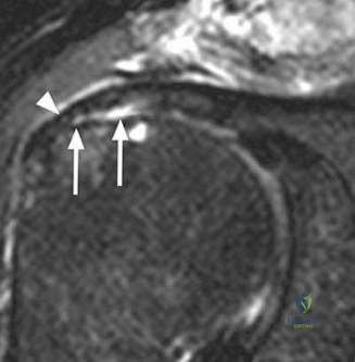
A 55-year-old active manual laborer presents to the emergency department following a fall onto his outstretched arm 3 weeks ago. He complains of persistent pain and significant weakness in shoulder abduction. Clinical examination reveals a positive Jobe’s test and external rotation lag sign. You obtain an MRI scan. Describe the critical prognostic indicators you are looking for on this image.
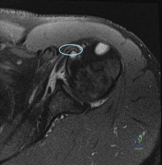
Candidate: I would look at the tear size and the quality of the muscle. I'll check for how far the tendon has retracted using the Patte classification and check for muscle atrophy and fatty infiltration using the Goutallier classification. I also want to see if the tear is reparable or if there is too much muscle degeneration.
Candidates often list classifications without explaining their clinical relevance. A poor answer ignores the distinction between "reparable" and "irreparable" anatomy, and fails to mention the importance of the humeral head position (e.g., superior migration suggesting chronicity or massive tear).
I would perform a systematic assessment of three key domains: 1. Tear Morphology: Assessing the size and the Patte classification of retraction. 2. Muscle Quality: Utilizing the Goutallier classification for fatty infiltration and looking for muscle belly atrophy on sagittal T1-weighted sequences. 3. Joint Integrity: Evaluating for superior humeral head migration, which indicates a loss of the rotator cuff force couple, and checking for associated pathology like biceps instability or labral injury. These factors collectively determine whether the tear is physiologically reparable or requires salvage reconstruction.
The MRI confirms a full-thickness, 2cm tear of the supraspinatus with minimal retraction. The patient is booked for an arthroscopic repair. Describe the biomechanical rationale for choosing a suture-bridge (transosseous equivalent) technique over a single-row repair.
Candidate: Suture-bridge repair is better because it provides a stronger construct. It spreads the tension across the whole footprint rather than just one line of anchors. Studies show it has higher load-to-failure strength and reduces gap formation, which helps the tendon heal better to the bone.
Failure to mention "footprint contact pressure" or "contact area." Simply stating it is "stronger" is not enough for an elite-level pass. Candidates should articulate the conversion of a point-loading force into a surface-area compression force.
The suture-bridge technique provides a transosseous equivalent repair. Its primary biomechanical advantage is the significant increase in footprint contact area and compressive force across the tendon-bone interface. By spreading the load across a medial and lateral row, it minimizes "gap formation" at the repair site during cyclic loading, facilitating superior biological healing by recreating the native enthesis footprint rather than a single-point fixation.
During your arthroscopic inspection, you note significant inflammation, but the patient's symptoms and history also raise a red flag for potential septic arthritis or crystal arthropathy. You see this image on your diagnostic sweep.
How do you manage this intra-operatively?
Candidate: I would stop the repair immediately. I’ll take samples for microbiology and histology (sent in sterile containers). I will perform a thorough lavage and debridement of the joint. I wouldn't place any hardware if there is clear pus, and I would start empirical antibiotics after consulting with microbiology.
Forgetting to mention the "histology" samples or failing to highlight the importance of "joint lavage" using large volumes of saline. Borderline candidates also fail to mention post-operative coordination with the infectious disease team.
Safety is the priority. 1. Cease the procedure to prevent further contamination. 2. Collect multiple samples (minimum of 3-5) for microscopy, culture, and sensitivity (MCS) and histopathology to rule out infection versus crystalline disease (e.g., gout/CPPD). 3. Aggressive Irrigation: Perform an extensive arthroscopic synovectomy and lavage with 10+ liters of saline. 4. Hardware decision: If the clinical suspicion for septic arthritis is high, I would not proceed with tendon repair or anchor placement. 5. Management: Close the portals, start targeted antibiotics only after formal consultation with Microbiology/ID, and discuss the plan for a staged reconstruction once the infection is cleared.
Detailed Chapters & Topics
Dive deeper into specialized chapters regarding shoulder-and-elbow-cases-acute-rotator-cuff-tears