Partial-Thickness Rotator Cuff Tears: Repair or Debride?
Key Takeaway
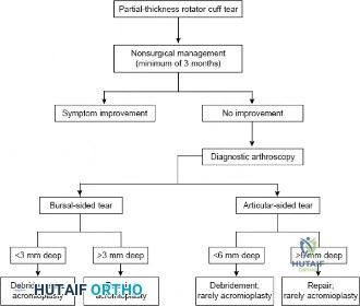
Your ultimate guide to Partial-Thickness Rotator Cuff Tears: Repair or Debride? starts here. For a 5mm deep partial-thickness rotator cuff tear, treatment is guided by the Ellman classification. An articular-sided tear (3-6mm) typically warrants debridement. Conversely, a bursal-sided partial-thickness rotator cuff tear of this depth (3-6mm) generally requires surgical repair. This ensures appropriate management based on tear characteristics.
You are evaluating a 45-year-old overhead athlete presenting with chronic, activity-related shoulder pain that has failed 4 months of structured physical therapy. You suspect a partial-thickness rotator cuff tear. How do you classify this pathology to guide your decision-making, and what specific anatomical considerations are critical for the surgical plan?
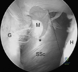
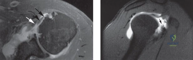
Candidate: I would use the Ellman classification, which categorizes tears by location (articular, bursal, or interstitial) and depth (Grade 1 <3mm, Grade 2 3-6mm, Grade 3 >6mm). Anatomically, I focus on the supraspinatus footprint, which is roughly 15mm wide. I need to distinguish between articular-sided (PASTA) lesions and bursal-sided lesions, as the latter are generally more symptomatic and prone to progression.
Candidates often focus solely on the classification without discussing the "50% rule" or the histology. A poor answer ignores the clinical significance of the "rotator cable," which maintains the mechanical integrity of the tendon crescent. Failing to mention why bursal-sided tears are more aggressive shows a lack of deep understanding.
The candidate should state: "I classify using the Ellman system. Crucially, I apply the '50% rule' (approx. 7-8mm of exposed footprint) to decide between debridement and repair. I must assess the rotator cable integrity; if the cable is compromised, repair is mandatory regardless of depth. Histologically, I recognize the 5-layer structure described by Clark and Harryman, noting that strain mismatch between the stiffer articular fibers and the more elastic bursal fibers explains the pathogenesis of articular-sided tears."
An MRI report suggests an articular-sided partial-thickness tear. You proceed to arthroscopy. How do you confirm the depth, and what is your decision-making process between an 'in-situ' transtendon repair versus tear completion and formal repair?
Candidate: I use a calibrated probe to measure the footprint distance from the articular margin. If >50%, I consider repair. Transtendon repair preserves the bursal fibers but is technically challenging and risks stiffness. Tear completion allows for a familiar anatomical repair but sacrifices healthy tissue.
Candidates often fail to mention "marker sutures" to correlate articular and subacromial findings. They also ignore the increased risk of postoperative stiffness, which is a major morbidity associated with transtendon repairs.
A structured answer: 1. Verification: Use a calibrated probe; if >7-8mm of footprint is exposed, it is a high-grade tear. Use a spinal needle to pass a marker suture into the subacromial space. 2. Transtendon: Pros: Preserves native bursal fibers; biomechanically sound. Cons: Risk of stiffness, "blind" knot tying. 3. Completion: Pros: Allows for anatomic footprint restoration and excision of degenerated tissue; lower risk of stiffness. Cons: Sacrifices potentially viable bursal tissue. 4. Conclusion: Both are clinically validated, but I prioritize the patient's stiffness risk and tissue quality when deciding.
You have performed a high-grade articular-sided repair. The patient is now 3 months post-op and reports significant stiffness, but the pain has improved. How do you manage this?
Candidate: I would diagnose secondary adhesive capsulitis. I would intensify the physical therapy program, focus on low-load prolonged stretching, and potentially offer a corticosteroid injection. If it persists beyond 6-9 months, I would consider an arthroscopic capsular release.
Immediately suggesting a Manipulation Under Anesthesia (MUA) or early surgical release without exhausting non-operative modalities or ensuring the tendon has matured sufficiently for the stress of manipulation.
The candidate should emphasize a conservative, stepwise approach: 1. Clinical Assessment: Exclude failure/retear (if in doubt, MRI). 2. Non-operative Management: Supervised, aggressive physical therapy (ROM-focused), education on the nature of post-surgical stiffness. 3. Pharmacology: Intra-articular steroid injections to reduce synovitis. 4. Surgical Escalation: Only after 6-9 months of failed conservative management, arthroscopic capsular release. Avoid MUA due to the risk of anchor pull-out in a healing repair.
