العربية
Comprehensive Master Guide · Medically Reviewed

Ponseti Clubfoot Correction: The Role of Percutaneous Achilles Tenotomy

Ponseti Casting DEFINITION Clubfoot, also known as congenital talipes equinovarus , occurs in approximately 1 in 1000 live births. The clubfoot contains four i…

246 Detailed Chapters
21 min read
Updated: مارس 2026
Dr. Mohammed Hutaif
Medically Reviewed by
Prof. Dr. Mohammed Hutaif
Verified Content Expert Reviewed

Quick Medical Answer

Learn more about Ponseti Clubfoot Correction: The Role of Percutaneous Achilles Tenotomy and how to manage it. Ponseti casting is the standard initial treatment for congenital talipes equinovarus (clubfoot), characterized by CAVE components and thickened Achilles tendons. This method uses serial casting to gently stretch soft tissues and remold cartilaginous bones. A percutaneous Achilles tenotomy is often performed as part of this process to fully correct the equinus deformity and achieve lasting correction.

Illustration of percutaneous achilles tenotomy - Dr. Mohammed Hutaif

Definition

Clubfoot, also known as congenital talipes equinovarus , occurs in approximately 1 in 1000 live births. The clubfoot contains four identifiable components that are easily remembered using the acronym CAVE (cavus, adductus, varus, and equinus). Idiopathic clubfoot contains each of the four components to varying degrees.

The so-called postural clubfoot is held by the infant in an equinovarus position, but all components are nearly completely correctable with gentle manipulation and resolve over time without intervention. A small proportion of clubfeet are teratologic, occurring as part of other neuromuscular diseases, such as Larsen syndrome, any of the arthrogryposis syndromes, and spina bifida. The complex clubfoot, a severe type of idiopathic clubfoot, has a tighter hindfoot and plantar structures.

In 1948, Dr. Ignacio Ponseti began manipulating clubfeet through serial casting, completely correcting the clubfoot deformity. The principles of Ponseti casting lay in gently stretching the soft tissue structures and gradually inducing remolding of the primarily cartilaginous bones of the hindfoot during immobilization. For the definitive publication on clubfoot and the Ponseti technique, the reader is referred to Dr. Ponseti's book. 7 The success of the treatment protocol that bears his name has been borne out through over 30 years of follow-up, establishing it as the standard for initial treatment of clubfoot. 1 In 2006, Dr. Ponseti published a modification to his original casting technique that addresses the specific deformities characteristic of the complex clubfoot. 8

Anatomy

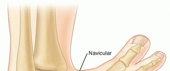
The Achilles and posterior tibialis tendons, as well as the posterior and medial ligaments of the foot between the calcaneus, talus, and navicular, are thickened and fibrotic. 7 The clubfoot contains a number of changes in bony alignment and shape.

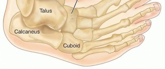
FIG 1: Anatomic alignment in neonatal clubfoot. Note the medial displacement of the navicular and cuboid, the inversion and internal rotation of the calcaneus under the talus, and equinus of the talus and calcaneus.

Relative to normal foot anatomy, the first ray is plantarflexed, generating the cavus deformity. By comparison, all rays are plantarflexed in the complex clubfoot, resulting in full-foot cavus. The navicular is medially displaced on the talus, and the cuboid is medially displaced on the calcaneus as part of the adductus deformity. The medial corners of the head of the talus and the anterior calcaneus are flattened. The calcaneus is inverted under the talus, creating the hindfoot varus, while also being in equinus and elevated in the fat pad of the heel.

In children with unilateral clubfoot, the affected foot usually is smaller, as is the lower leg, relative to the unaffected side. Up to 85% of clubfeet have an insufficient or absent anterior tibial artery. 6

Natural History

The exact cause of the fibrotic changes in clubfoot is unknown. Recently, candidate genes have been identified in familial clubfoot, including Pitx1 and Tbx4 . 3 Left uncorrected, the weight-bearing surface in a clubfoot becomes the dorsolateral surface. Thick callosities develop, and the positioning of the foot creates significant functional disability.

Patient History and Physical Findings

Clubfoot may be identified on prenatal ultrasound as early as 12 to 13 weeks. Half or more of fetuses with clubfeet identified on second-trimester ultrasounds are found to have other anomalies (most commonly cardiac, neurologic, and/or urogenital) or are syndromic/teratologic. 9 The exact sensitivity and specificity of prenatal ultrasound are unknown. False positives are rare on 20-week ultrasounds but may be as high as 40% during the third trimester (when false negatives are rare). 9 Cases not found on prenatal ultrasound are readily identifiable at birth.

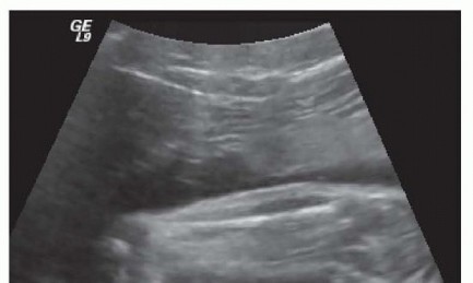
FIG 2: Ultrasound at 20 weeks of a child born with clubfoot.

All children with clubfeet should be examined for other findings that may suggest a syndromic or neuromuscular association, such as other contractures or joint dislocations (especially hip dislocation), cutaneous lesions, spinal abnormalities, and abnormal facial features. The clubfoot is easily identified by the combined deformities of cavus, adductus, varus, and equinus.

Consider complex clubfoot if a deep midfoot crease and cavus extend transversely across the entire plantar aspect of the foot, and the foot appears short and broad. The ability to abduct or dorsiflex the foot completely on examination suggests etiologies other than idiopathic clubfoot, such as isolated metatarsus adductus, neuromuscular disease, or focal anatomic abnormalities.

The fat pad of the heel will feel empty upon palpation due to equinus positioning of the calcaneus. This is especially dramatic in the complex clubfoot. The lateral head of the talus is easily palpable over the dorsolateral surface of the foot. More laterally, the anterior calcaneal tuberosity is also palpable. Care must be taken in differentiating these two structures because Ponseti casting necessitates stabilizing the foot over the lateral head of the talus, allowing free motion of the calcaneus under the talus, whereas pressure at the calcaneal tuberosity blocks calcaneal rotation, allowing only forefoot abduction.

The complex clubfoot has a crease that extends transversely across almost the entire plantar aspect of the foot accompanied by full-foot cavus with plantarflexion of all metatarsals. Also, the heel crease is deeper than that of most other clubfeet. During the initial one or two casts, as the adductus is corrected, the first ray in the complex clubfoot becomes retracted, if not noticeably retracted at presentation. The cavus also persists, with all metatarsals remaining plantarflexed.

It is important to examine the clubfoot before each casting to evaluate for the adjustments that must be made during casting to correct residual deformities or to identify and modify casting for a complex clubfoot. A number of classification systems have been introduced as an attempt to predict outcome, but the ability of these systems to evaluate correction, predict recurrence and final function is still unclear. 5 The degree of dorsiflexion and abduction, and the distance of the navicular anterior to the medial malleolus, provide other objective measurements of deformity and correction.

Some children are born with one or both feet held in an equinovarus deformity at birth that is nearly completely correctable on examination. Nearly complete dorsiflexion (more than 20 degrees) is present, although abduction may be slightly limited. The calcaneus is also readily palpable in the fat pad of the heel. These feet may be thought of as "postural" in nature, and most will resolve spontaneously or with parental stretching over 1 to 2 months. If persistent, one or two casts usually correct the deformity, and Achilles tenotomy is rarely required. Feet corrected with casting may require maintenance in a foot abduction orthosis.

Imaging and Other Diagnostic Studies

Clinical examination is sufficient to diagnose the congenital clubfoot. Plain radiographs at birth are not helpful in diagnosing clubfoot because the ossific nuclei of the talus and calcaneus are spherical, so orientation and relationship are not discernible, and the other tarsal bones are unossified.

Once full abduction is obtained by casting, if dorsiflexion of more than 10 degrees is present, forced dorsiflexion lateral films are helpful in differentiating midfoot breach, producing apparent dorsiflexion, from true dorsiflexion occurring at the ankle, obviating the need for a percutaneous Achilles tenotomy.

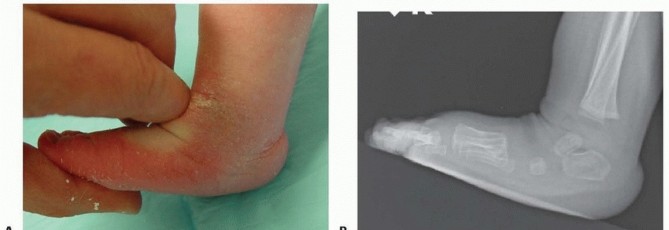
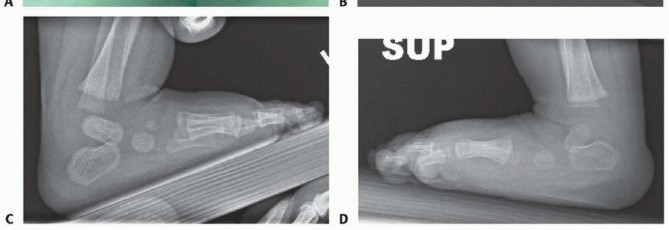
FIG 3: Forced dorsiflexion lateral radiographs to differentiate midfoot breach from true ankle dorsiflexion.

Differential Diagnosis

  • Metatarsus adductus
  • Neurologic equinovarus or cavovarus deformity: Both deformities may be differentiated from clubfoot by absence of the other components of clubfoot.
  • Teratologic or syndromic clubfeet (including neuromuscular disorders): Clubfoot deformity may be more difficult to correct and tends to recur.
  • Postural clubfoot
  • Complex clubfoot

Nonoperative Management

Ponseti casting of the idiopathic clubfoot involves a specific sequence of corrective maneuvers that correct the deformities of the clubfoot in combination. Each manipulation is maintained with a plaster cast. Ponseti casting ideally begins during infancy, although good results are achievable through toddlerhood. Casting in older children can also produce good results or at least reduce the amount of surgery required for complete correction. An open tendo Achilles lengthening may be more appropriate than a percutaneous tenotomy in children older than 2 years old.

Long-leg casts should always be used to prevent cast slippage and maintain rotational control of the lower leg. Initially, applying a short-leg cast allows focused attention on maintaining foot position and molding before extending the cast above the knee. Padding should be minimal, and plaster is preferable for its ability to be molded precisely to the contours of the foot and ankle.

Four to six casts should correct the cavus, adductus, and varus deformities. If correction is not achieved in eight casts or the child pulls back in the casts, the possibility of an unrecognized complex clubfoot or improper casting technique should be considered. Casting is facilitated by the child being relaxed and calm. Feeding the infant during casting assists in this. For breast-fed infants, it is helpful if the family introduces, and uses once daily, a bottle so the child may feed during casting. If a bottle is not tolerated, other calming measures may be necessary. For older children, music, television, or playing with toys often proves helpful, as does casting with the child sitting upright or on the parent's lap.

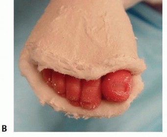
**Vascular Check:** Before leaving the clinic, the toes should be checked to make sure they are pink and well perfused. Some toes will become reddish-purple as the casts cool but will become pink if the child is bundled and monitored over 1 hour. Toes that become more purple and dusky indicate that the cast is too tight and should be reapplied.

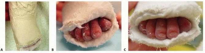
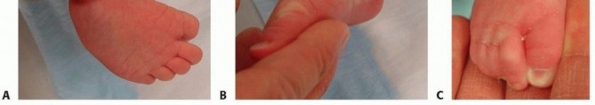
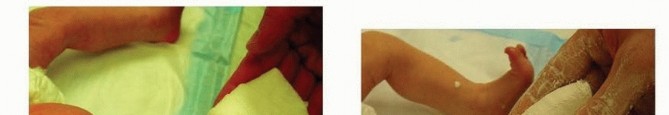
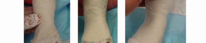
FIG 4: (A) A complex clubfoot that has pulled back in the cast. (B) Purple discoloration of toes after application. (C) Reperfusion and pink toes after the cast temperature stabilizes.

Casts are changed every 5 to 7 days. The final cast, following percutaneous Achilles tenotomy, is left in place for 3 weeks. Almost all clubfeet will require a percutaneous Achilles tenotomy to correct the residual equinus deformity once the other components are corrected. Once complete correction is obtained, correction must be maintained by placing the feet in a foot abduction orthosis. Constructs include straight last shoes, soft ankle-foot orthoses (AFO), or rigid AFOs mounted on rigid or articulating bars.

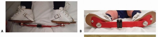
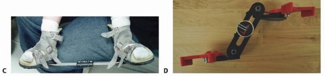
FIG 5: The foot abduction orthosis constructs (straight last shoes attached to a solid bar, and soft AFO options).

Surgical Management

Percutaneous Achilles tenotomy is required in almost all idiopathic clubfeet to correct the residual equinus. About 20% of patients require anterior tibialis tendon transfer at 3 to 4 years old to correct recurrent or persistent dynamic varus deformity.

Preoperative Planning

  • Degree of dorsiflexion: If dorsiflexion is less than 10 degrees, a percutaneous Achilles tenotomy is required to correct the residual equinus. If dorsiflexion is more than 10 degrees, forced dorsiflexion lateral foot radiographs help to differentiate midfoot dorsiflexion, with residual calcaneal equinus, from true dorsiflexion occurring at the hindfoot.
  • Location & Anesthesia: The risk of anesthesia must be balanced against the perceived pain and duration of the procedure. Approximately half of pediatric orthopedists report performing the percutaneous tenotomy under general anesthesia or conscious sedation. Local analgesia, with 1% lidocaine, affords the opportunity to perform the tenotomy in the clinic setting and avoids any potential risk of general anesthesia.

Positioning

The child should be supine on the table with the contralateral leg held out of the way by the parent or an assistant during tenotomy and casting.

Approach

A medial approach is used to remain posterior to the medial neurovascular bundle.


Techniques

1. Casting

Stretching
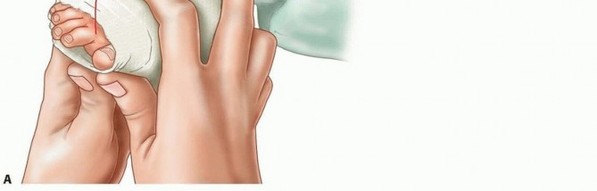
Before casting, the foot should be stretched in the same manner as used for immobilization during casting. The thumb of the examiner's contralateral hand should be placed over the head of the talus, and the index finger of the other hand should lie along the medial aspect of the first ray with the second through fourth fingers under the plantar aspects of the forefoot. The calcaneocuboid joint should be avoided, so as not to block subtalar motion. The first casting should focus on elevation of the first ray to correct the cavus deformity. This places the forefoot in supination, locking the midfoot and aligning the forefoot with the hindfoot, allowing for correction of the hindfoot deformities during later abduction maneuvers. Some of the adductus may also be corrected during the first casting.

TECH FIG 1: Proper hand placement during stretching and first cast application for elevation of the first ray.

Lower Leg Cast Application
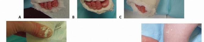
A thin layer of cotton padding should be applied. The padding is wrapped three times around the toes distally, then extended proximally over the foot and lower leg to pad with no more than two layers of padding. The foot should be held in the position to be casted throughout. The popliteal fossa should be avoided proximally. A thin layer of plaster is applied over the foot and lower leg. The plaster may be applied more loosely over the toes but should be snug over the hindfoot and ankle to immobilize the foot properly and allow for precise molding.

Avoid making the cast too snug so as to impair venous return or apply unnecessary pressure on the fat pad of the heel. The lower leg cast should be precisely molded around the malleoli and above the calcaneus posteriorly. Do not apply pressure over the fat pad of the heel. Throughout, the foot should be held in the position of correction, but the fingers should be in fairly constant motion to prevent pressure spots.

TECH FIG 2: Holding the position of correction while applying thin plaster and molding around the malleoli.

Completing the Cast
Once the lower leg cast has set, padding should be applied over the rest of the leg up to the groin, again in no more than two or three layers. The knee should be held at 90 degrees, and the lower leg should be in slight external rotation. Padding should be minimized in the popliteal fossa to prevent impingement of the neurovascular structures. A plaster splint of three or four layers of plaster roll should be placed over the knee from the proximal thigh to the middle of the shin to strengthen the cast against knee extension while minimizing bulk in the popliteal fossa.

The knee should be molded while held at 90 degrees with the lower leg in slight external rotation until set. Rolling the plaster at the proximal edge of the cast before the plaster sets up completely helps minimize chafing of the thigh.

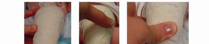
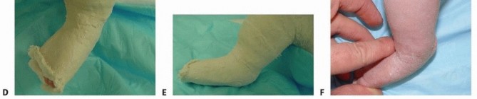
TECH FIG 3: Extending the cast to a long-leg cast and trimming the distal end to ensure pink, well-perfused digits.

The cast should be trimmed distally to expose the toes. The practitioner should confirm that they are pink and well perfused before the child is sent home. Trimming the plaster over the dorsal aspect too far proximally, beyond the web space, may create a tourniquet effect over the forefoot. Parents should be instructed on signs and symptoms of cast problems before discharge.

Cast Changes and Follow-Up
Casts are typically changed every 7 days, although they may be changed as frequently as every 5 days. Casts should not be removed until just before recasting. Casts can be soaked by the family before coming to the office, then removed with a plaster knife in the clinic. Alternatively, dry casts may be removed with a cast saw, using extreme caution. Having the parents remove the casts the night before results in varied degrees of recurrence overnight and prolongs casting.

After the first casting, the cavus deformity should be nearly, or completely, corrected. If not, adopt complex casting modifications. Stretching is performed with the forefoot in supination, maintaining alignment of all rays, abducting the foot under the talus, again stabilizing the talus laterally. The foot is then casted in the newly maintained position, just to where the foot may be comfortably corrected without significant resistance. Trying to overabduct the foot during a single casting results in intolerance, pressure sores, or vascular compromise.

Each subsequent manipulation results in increased abduction of the forefoot and correction of the hindfoot varus. Throughout, the forefoot should remain in neutral (appearing supinated due to the hindfoot varus) and the hindfoot in equinus. Dorsiflexion of the calcaneus remains blocked under the neck of the talus until approximately 25 degrees of abduction has been obtained. Once abduction of 70 degrees is obtained, correction of the remaining equinus deformity may occur. Overabduction to 70 degrees is necessary to accommodate some of the inevitable recurrence.

TECH FIG 4: Progression of correction through subsequent casts, decreasing varus and equinus and increasing abduction to 70 degrees.

2. Complex Clubfoot Casting

The complex clubfoot may not be immediately recognizable at presentation. Correction usually begins using the standard maneuvers, but within one or two casts, the foot begins to clearly demonstrate a deviation from the expected correction as the cavus persists (involving plantarflexion of all metatarsals) and the first ray becomes retracted. At this point, the technique must be modified.

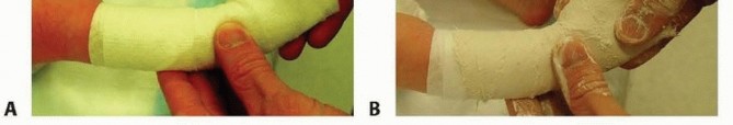
Lateral counterpressure still occurs at the lateral head and neck of the talus, but stabilization of the fibula should also occur. The index finger of the contralateral hand should be flexed at the proximal interphalangeal joint and placed posterior to the distal fibula. The thumb of the same hand is placed just anterior to the lateral malleolus along the neck of the talus.

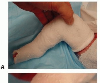
After applying cotton padding, a posterior splint of three or four layers of plaster should be applied under the plantar surface of the foot, extending from beyond the tips of the toes proximally over the posterior lower leg. Then, a thin layer of plaster may be wrapped in the usual manner. A minimal amount of plaster should be used because precise molding is even more important for the complex clubfoot.

The pads of the thumbs of both hands are placed under the forefoot, with the pads of the index fingers placed over the dorsal surface of the talar neck. The forefoot is then forcefully dorsiflexed against the counterpressure over the dorsal talar neck, enough to produce blanching of the digits. Upon release of dorsiflexion pressure after setting of the cast, the slight relaxation of the cast should result in revascularization of the digits and pink coloration.

TECH FIG 5: Dorsiflexion force applied to stretch the midfoot in the complex clubfoot, checking for digit blanching and reperfusion.

On extending the cast up over the lower leg, the knee should be flexed to 110 degrees to minimize the ability to pull out of the cast. Tenotomy occurs once the cavus and adductus deformities are corrected and about 40 degrees of abduction is obtained. Attempting to abduct the complex clubfoot beyond 40 degrees results in no further hindfoot correction and only overabducts the forefoot.

3. Percutaneous Achilles Tenotomy

The tenotomy should occur 1 to 1.5 cm above the insertion of the Achilles on the posterior tuberosity of the calcaneus. Performing the tenotomy too low results in damage to the posterior calcaneal tuberosity. For procedures in the clinic, local anesthesia must be used. A small amount of 1% lidocaine may be injected locally adjacent to the tendon.

An assistant should hold the foot in maximal dorsiflexion to increase tension on the Achilles tendon, making it more easily palpable. A second assistant should hold the contralateral leg and foot out of the field. A thin, sharp scalpel should be used to perform the tenotomy. Cataract surgical blades (5100 or 5400 Beaver blades) are well suited for this procedure, although a no. 11 blade is also acceptable.

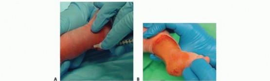
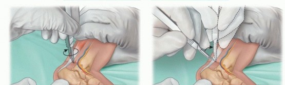
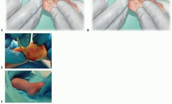
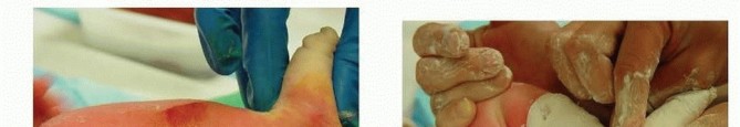
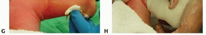
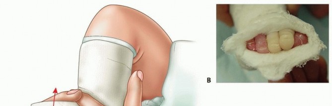
TECH FIG 6: Step-by-step percutaneous Achilles tenotomy under local anesthesia, blade positioning, transection, and post-tenotomy cast molding.

Once the blade is oriented perpendicular to the fibers of the Achilles tendon, the safest maneuver involves pressing the tendon onto the blade using the contralateral thumb. Complete transection often results in a palpable "pop," release of the Achilles tendon, and an immediate increase of 15 to 20 degrees of dorsiflexion. A palpable defect in the tendon confirms complete transection.

Casting After Tenotomy: The lower leg is wrapped with sterile cotton, accommodating the increased dorsiflexion. The plaster is applied in the usual manner and molded well at the anterior ankle to prevent pulling back in the cast. Extension of the cast above the thigh as a long-leg cast should occur with the knee in the usual 90 degrees of flexion (110 degrees for complex clubfoot). The post-tenotomy cast should be left on for 3 weeks before removal to allow tendon healing.

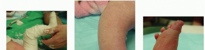
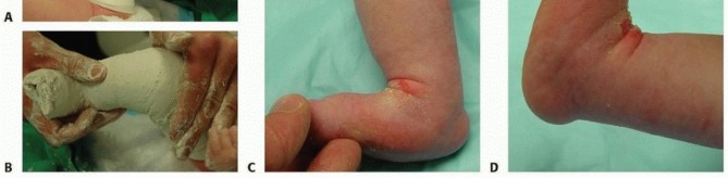
TECH FIG 7: Final long-leg casting and complete correction obtained three weeks post-tenotomy.

Pearls and Pitfalls

  • Failure to correct the cavus deformity with initial casting: Failure to elevate the first ray will result in worsening cavus during abduction, and only the forefoot will abduct. The hindfoot varus will fail to correct. The foot will then pull back in the cast.
  • Toes turn purple after cast application (see FIG 4): Some neonatal feet have poor vascular control and will turn purple as the cast cools. Do not be too hasty to remove the cast. Bundle the child, elevate the feet, and recheck every 15 minutes. Increasing purplish discoloration indicates a cast that is too tight and should be removed and reapplied.
  • An older child who resists casting: A child who fights casting prevents good molding, and too much motion may prevent the cast from setting up in the desired position. A quiet room with music, entertaining the child with a toy, or feeding may relax the child.
  • Child pulling out of foot abduction orthosis: Add padding in the heel, above the posterior calcaneal tuberosity, use a shoe with a heel cutout, or both. If the child has a strong propensity for toe curling, try a Plastazote plate under the toes to keep them extended.
  • Child cries while in casts or in bar and shoes: Make sure the toes are well perfused. Discomfort for 24 hours after the first casting or tenotomy is common and easily relieved with acetaminophen. If the child is in an orthosis, examine the feet for sores. Feet may be hyperesthetic after casting: massage during diaper changes accelerates desensitization.
  • Recurrence: Monitor for decreases in abduction and dorsiflexion. Treating an early identified, minimal recurrence with stretching by the parents with every diaper change may prevent progression. Later or more marked recurrence should be treated with recasting and possibly a second percutaneous tenotomy.

Postoperative Care

After removal of the post-tenotomy cast, the child should immediately be placed in a foot abduction orthosis. Acceptable constructs include straight last shoes or AFOs connected to a solid or articulated bar.

In the case of bilateral corrected clubfeet, both shoes should be placed in abduction/external rotation on the bar to the degree of comfortable correction, typically 60 to 70 degrees. For unilateral clubfoot, only the shoe of the affected foot is placed near the extreme of abduction; the shoe of the normal foot is placed at 30 degrees.

Only a single, thin pair of socks should be worn with the shoes. For the first week, the orthosis and socks should be removed with every diaper change to inspect the feet for evidence of developing pressure sores. Red spots that do not disappear within 5 minutes signal a potential problem spot and require refitting of the shoes with Plastazote or repositioning on the bar.

After the first week, the orthosis should be worn full time, but it may be removed once daily for bathing and a short period of play (1 to 2 hours). Full-time wear continues for 3 to 4 months to maintain correction. After 3 months of full-time wear and maintenance of full correction, children wear the orthosis for 16 hours per day, primarily at nighttime and during naps. Part-time wear continues until the child is 4 years old, when orthosis wear may be discontinued.

Outcomes

A corrected clubfoot tends to recur to its original position, requiring maintenance of correction in the orthosis. Noncompliance with bar-and-shoe wear increases the likelihood of recurrence to more than 80%. Compliance is increased with close follow-up and explicit discussions with the family and all caregivers. 4 Twenty percent to 50% of corrected clubfeet will require anterior tibialis tendon transfer to correct dynamic varus present during ambulation.

Complications

  • Cast sores, cast saw burns.
  • Prolonged casting or pulling back in the cast due to improper technique, unrecognized clubfoot, or failure to modify casting for complex clubfoot.
  • Overabduction from unrecognized complex clubfoot or overabduction in foot abduction orthosis (beyond degree of correction).
  • Posterior tibial artery impingement.
  • Peroneal artery or lesser saphenous vein laceration during tenotomy. 2
  • Recurrence due to incomplete correction or lack of orthosis wear.

Scientific References

  1. Cooper DM, Dietz FR. Treatment of idiopathic clubfoot. A thirty-year follow-up note. J Bone Joint Surg Am 1995;77(10):1477-1489.
  2. Dobbs MB, Gordon JE, Walton T, et al. Bleeding complications following percutaneous tendo Achilles tenotomy in the treatment of clubfoot deformity. J Pediatr Orthop 2004;24:353-357.
  3. Dobbs MB, Gurnett CA. Genetics of clubfoot. J Pediatr Orthop B 2012;21:7-9.
  4. Dobbs MB, Rudzki JR, Purcell DB, et al. Factors predictive of outcome after use of the Ponseti method for the treatment of idiopathic clubfeet. J Bone Joint Surg Am 2004;86-A(1):22-27.
  5. Flynn JM, Donohoe M, Mackenzie WG. An independent assessment of two clubfoot-classification systems. J Pediatr Orthop 1998;18: 323-327.
  6. Greider TD, Siff SJ, Gerson P, et al. Arteriography in club foot. J Bone Joint Surg Am 1982;64(6):837-840.
  7. Ponseti IV. Congenital Clubfoot: Fundamentals of Treatment. New York: Oxford University Press, 1996.
  8. Ponseti IV, Zhivkov M, Davis N, et al. Treatment of the complex idiopathic clubfoot. Clin Orthop Relat Res 2006;451:171-176.
  9. Treadwell MC, Stanitski CL, King M. Prenatal sonographic diagnosis of clubfoot: implications for patient counseling. J Pediatr Orthop 1999;19:8-10.
  10. Zionts LE, Sangiorgio SN, Ebramzadeh E, et al. The current management of idiopathic clubfoot revisited: results of a survey of the POSNA membership. J Pediatr Orthop 2012;32:515-520. [View Source / PubMed]

Detailed Chapters & Topics

Dive deeper into specialized chapters regarding ponseti-casting

246 Chapters
01
Chapter 1 18 min

Midshaft Clavicle Fractures: An Evidence-Based Guide to Diagnosis, Management, and Outcomes

A comprehensive guide to midshaft clavicle fractures, detailing epidemiology, anatomy, biomechanics, and evidence-based…

02
Chapter 2 26 min

Proximal Humerus Fractures Epidemiology: Trends, Causes & Management

Learn about proximal humerus fractures, a common type of fracture often seen in older patients with osteoporotic bones.…

03
Chapter 3 29 min

Scapula Fractures: Epidemiology, Classification, Anatomy, and Management

Scapula fractures are rare but signify high-energy trauma, mandating thorough evaluation for associated injuries. Disco…

04
Chapter 4 22 min

Proximal Humerus Fractures: Essential Etiology, Diagnosis, and Treatment

Learn about proximal humerus fractures, their prevalence, causes, diagnostic methods, and treatment options. Find infor…

05
Chapter 5 7 min

Scapula Fractures: Decoding the Causes, Diagnosis & Treatment

Scapula Fractures: Causes, Diagnosis and Treatment Scapula Fractures: Causes, Diagnosis and Treatment Summary Scapula F…

06
Chapter 6 24 min

Humerus Fractures: A Comprehensive Surgical Review for Orthopedic Professionals

Explore a comprehensive surgical review of humerus fractures, covering proximal, diaphyseal, and distal types. Delve in…

07
Chapter 7 32 min

Distal Clavicle Fractures: Essential Guide to Diagnosis & Treatment

Distal Clavicle Fractures: Diagnosis, Treatment, and Complications Distal Clavicle Fractures Distal clavicle fractures …

08
Chapter 8 20 min

Superior Glenohumeral Dislocation: A Comprehensive Guide to Diagnosis, Surgical Anatomy, and Management

Superior glenohumeral dislocation is a rare, complex shoulder injury. Learn about its diagnosis, surgical anatomy, biom…

09
Chapter 9 21 min

Superior Glenohumeral Dislocation Mechanism: Injury to Recovery Guide

Superior Glenohumeral Dislocation: Mechanism of Injury, Evaluation, Treatment, and Complications Superior Glenohumeral …

10
Chapter 10 12 min

Heal Your AC Joint Injury: Causes, Symptoms & Treatment Guide

Acromioclavicular (AC) joint injury is a common shoulder injury that occurs when the ligaments that connect the collarb…

11
Chapter 11 8 min

Clavicle Fractures: Epidemiology, Anatomy & Treatment Insights

Clavicle fractures are common injuries that affect the collarbone, which connects the shoulder to the chest. They can c…

12
Chapter 12 32 min

Orthopaedic Problems in Children with CP: Prevention & Treatment

This blog post discusses the most common orthopaedic problems in children with cerebral palsy, as well as how to preven…

13
Chapter 13 8 min

Cerebral Palsy: Orthopaedic Foot & Ankle Solutions

Cerebral palsy (CP) is a group of conditions that involve permanent motor dysfunction due to abnormalities of the devel…

14
Chapter 14 32 min

Mastering Orthopaedic Management of Cerebral Palsy

This article provides a comprehensive overview of orthopaedic management of cerebral palsy, including assessment and ev…

15
Chapter 15 30 min

Humerus Fractures: Surgical Anatomy, Epidemiology, & Management

Understand humerus fractures: epidemiology (proximal, diaphyseal, distal), detailed surgical anatomy, critical neurovas…

16
Chapter 16 26 min

Humerus Fractures: A Comprehensive Orthopedic Guide to Epidemiology & Anatomy

Dive into a comprehensive guide on humerus fractures, exploring their epidemiology, surgical anatomy of proximal, diaph…

17
Chapter 17 13 min

Pediatric Septic Hip: Know the Signs, Save the Joint

Pediatric septic hip arthritis is a serious condition that occurs when bacteria infect the hip joint of a child. It can…

18
Chapter 18 25 min

Unlocking Better Foot Function: Anterior Tibialis Transfer for Clubfoot

Clubfoot is one of the most common birth defects that affects the foot and ankle. It causes the foot to point downward …

19
Chapter 19 27 min

Congenital Foot Anomalies: Epidemiology, Anatomy & Pathoanatomy Review

Explore congenital foot anomalies: types (clubfoot, metatarsus adductus, CVT), epidemiology, and detailed pathoanatomy.…

20
Chapter 20 11 min

Demystifying Shoulder Girdle Anatomy: Bones, Joints & Movement

The shoulder girdle is a complex joint that connects the upper extremity to the axial skeleton. It is composed of four …

21
Chapter 21 40 min

Mastering Shoulder Anatomy: An Interactive Orthopedic Guide

ANATOMICAL FEATURES Fig. 4.A. The shoulder is complex, and it is important to note that it has two main components, nam…

22
Chapter 22 62 min

Dislocation of the Hip: Interactive Guide to Understanding DDH

DEVELOPMENTAL DISLOCATION OF THE HIP (DDH) This condition occurs in the perinatal period and involves displacement of t…

23
Chapter 23 30 min

Medial Clavicular Fragment Fractures: Risks, Symptoms, & Recovery

Clavicle Fractures EPIDEMIOLOGY Clavicle fractures account for approximately 2.6% of all fractures and for 44% to 66% o…

24
Chapter 24 13 min

Shoulder Ultrasound: Patient & Probe for Efficient, Precise Scans

Shoulder ultrasound is a safe, efficient, and cost-effective method for diagnosing rotator cuff tears. It is comparable…

25
Chapter 25 25 min

Shoulder-Spine Syndrome: Comprehensive Diagnosis & Management

Shoulder-Spine Syndrome presents diagnostic challenges with overlapping neck and shoulder pain. Understand its epidemio…

26
Chapter 26 31 min

Advanced Perioperative Pain Management for Shoulder Surgery: Regional Anesthesia & Neuroanatomy

Optimize perioperative pain for shoulder surgery. This guide details regional anesthesia, PNBs, and multimodal strategi…

27
Chapter 27 17 min

Magnetic Resonance Imaging in Shoulder Pathology: Diagnosis & Surgical Insights

Explore the critical role of MRI in diagnosing complex shoulder pathologies like rotator cuff tears, labral lesions, an…

28
Chapter 28 17 min

Understanding & Treating Glenohumeral Ligament HAGL Tears

Background, Diagnosis, and Interpretation Injuries to the inferior glenohumeral ligament (IGHL) and capsulolabral compl…

29
Chapter 29 12 min

Optimizing Perioperative Pain Management in Shoulder Surgery: An Academic Review

Explore advanced perioperative pain management for shoulder surgery, emphasizing regional anesthesia benefits. Learn ab…

30
Chapter 30 8 min

Complications in Arthroscopic Shoulder Surgery: What You Must Know

Summary Arthroscopic shoulder surgery complications are considered rare occurrences. However, there have been reports i…

31
Chapter 31 19 min

Acromioclavicular (AC) Joint Injuries: Epidemiology, Anatomy, Rockwood Classification & Management

Understand Acromioclavicular (AC) joint injuries: epidemiology, Rockwood classification (Types I-VI), detailed surgical…

32
Chapter 32 22 min

Midshaft Humerus Fractures: Epidemiology, Surgical Anatomy, Biomechanics & Management

Understand midshaft humerus fractures: epidemiology, detailed surgical anatomy (radial nerve risk), biomechanics of fix…

33
Chapter 33 25 min

Anterior Glenohumeral Dislocation: Comprehensive Guide to Epidemiology, Anatomy, Biomechanics & Management

Explore anterior glenohumeral dislocation: its epidemiology, injury mechanisms, and critical surgical anatomy. Understa…

34
Chapter 34 19 min

Posterior Glenohumeral Dislocation: Epidemiology, Anatomy, Diagnosis & Management

Understand posterior glenohumeral dislocation (PGHD): epidemiology, mechanisms (seizures, electric shock), key anatomy …

35
Chapter 35 23 min

Anterior Shoulder Dislocation: Epidemiology, Pathoanatomy, Diagnosis & Management

Understand anterior shoulder dislocation epidemiology, high recurrence, & critical associated injuries like Bankart & H…

36
Chapter 36 21 min

Clavicle Fracture: Comprehensive Orthopedic Guide to Epidemiology, Anatomy & Management

Understand clavicle fracture epidemiology, surgical anatomy, biomechanics, and indications for operative vs. non-operat…

37
Chapter 37 18 min

Mastering Femoral Head Autograft in DDH Hip Reconstruction

INDICATIONS Total hip replacement (THR) in the setting of developmental dysplasia of the hip (DDH) is indicated in symp…

38
Chapter 38 22 min

Proximal Humerus Fractures: Epidemiology, Classification, Anatomy & Management

Delve into proximal humerus fractures: epidemiology, Neer classification, and critical surgical anatomy, including vasc…

39
Chapter 39 11 min

Solving Unstable SCFE: Subcapital Realignment with Extended Retinacular Flap

Demographics Age : 12 years Sex : Female BMI : 19 Relevant Past Medical History Principal pathologies : Inconspicuous a…

40
Chapter 40 32 min

Pediatric Knee Fractures: When is a Long Leg Cast Necessary?

PEDIATRIC KNEE OVERVIEW The knee is a ginglymoid (hinge) joint consisting of three articulations: patellofemoral, tibio…

41
Chapter 41 7 min

Heal Your Clavicle Fractures: Expert Guide & Recovery Tips

CLAVICLE FRACTURES EPIDEMIOLOGY Clavicle fractures account for approximately 2.6% of all fractures and for 44% to 66% o…

42
Chapter 42 26 min

Posterior Glenohumeral Dislocation: Pathophysiology, Diagnosis, and Management

Posterior glenohumeral dislocation is frequently missed. Learn its epidemiology, mechanisms (seizures, trauma), subtle …

43
Chapter 43 8 min

Pediatric Hip Fractures: What Every Clinician Must Know

PEDIATRIC HIP PEDIATRIC HIP FRACTURES Epidemiology Hip fractures are rare in children, occurring less than 1% as often …

44
Chapter 44 36 min

Luxatio Erecta (Inferior Glenohumeral Dislocation): Anatomy, Diagnosis & Management

Explore Luxatio Erecta, a rare inferior glenohumeral dislocation. Understand its unique pathomechanics, high neurovascu…

45
Chapter 45 16 min

Glenohumeral Dislocation: Comprehensive Guide to Shoulder Stability

GLENOHUMERAL DISLOCATION EPIDEMIOLOGY The shoulder is the most commonly dislocated major joint of the body, accounting …

46
Chapter 46 9 min

Scapula Fractures: Your Guide to Recognizing and Treating Scapula Injuries

SCAPULA FRACTURES EPIDEMIOLOGY This relatively uncommon injury represents 3% to 5% of shoulder fractures and 0.4% to 1%…

47
Chapter 47 9 min

AC Joint Injury? Effective Healing with a Clavicle Strap Sling

ACROMIOCLAVICULAR AND STERNOCLAVICULAR JOINT INJURIES ACROMIOCLAVICULAR JOINT INJURY Epidemiology It is most common in …

48
Chapter 48 25 min

Children's Wrist and Hand Fractures: What You Need to Know

PEDIATRIC WRIST AND HAND INJURIES TO THE CARPUS Epidemiology Rare, although carpal injuries may be underappreciated owi…

49
Chapter 49 19 min

Pediatric Tibia and Fibula Fractures: Essential Clinical Insights

PEDIATRIC TIBIA AND FIBULA EPIDEMIOLOGY Tibia fractures represent the third most common pediatric long bone fracture, a…

50
Chapter 50 64 min

Pediatric Elbow Fractures & Dislocations: What You Need to Know

PEDIATRIC ELBOW EPIDEMIOLOGY Elbow fractures represent 8% to 9% of all upper extremity fractures in children. Of all el…

51
Chapter 51 38 min

Superior Glenohumeral Dislocation: Epidemiology, Anatomy, and Associated Injuries Guide

Explore superior glenohumeral dislocation, a rare, complex injury with extensive osseous and soft tissue damage. Unders…

52
Chapter 52 40 min

Master Pediatric Femoral Shaft & Knee Fractures: Interactive Module

PEDIATRIC FEMORAL SHAFT EPIDEMIOLOGY Represent 1.6% of all fractures in the pediatric population. Males are more common…

53
Chapter 53 31 min

Unraveling Pediatric Shoulder Fractures & Dislocations (Ages 0-18)

PEDIATRIC SHOULDER PROXIMAL HUMERUS FRACTURES Epidemiology These account for <5% of fractures in children. Incidence ra…

54
Chapter 54 14 min

Pediatric Forearm Fractures: Your Guide to Long Arm Casts

PEDIATRIC FOREARM EPIDEMIOLOGY These injuries are very common. They make up 40% of all pediatric fractures (only 4% are…

55
Chapter 55 28 min

Pediatric Ankle & Foot Fractures: Understanding Short Leg Walking

PEDIATRIC ANKLE EPIDEMIOLOGY Ankle injuries account for up to 18% of all physeal injuries; they are third in frequency …

56
Chapter 56 10 min

Proximal Humerus Fractures: Essential Guide to Anatomy & Recovery

PROXIMAL HUMERUS FRACTURES EPIDEMIOLOGY Proximal humerus fractures comprise 4% to 5% of all fractures and represent the…

57
Chapter 57 16 min

Distal Humerus Fractures: Understanding Causes, Anatomy & Epidemiology

DISTAL HUMERUS fractures EPIDEMIOLOGY This is a relatively uncommon injury, comprising approximately 2% of all fracture…

58
Chapter 58 11 min

Mastering Pediatric Fractures and Dislocations: Essential Principles

PEDIATRIC ORTHOPAEDIC SURGERY: GENERAL PRINCIPLES OVERVIEW The development and growth of the skeletal system from gesta…

59
Chapter 59 27 min

Anterior Glenohumeral Dislocation: Comprehensive Guide to Epidemiology, Anatomy, & Mechanisms

Delve into anterior glenohumeral dislocation, covering its epidemiology, detailed surgical anatomy including static/dyn…

60
Chapter 60 23 min

Proximal Humerus Fractures: Epidemiology, Advanced Anatomy & Biomechanics

Understand proximal humerus fractures: their bimodal epidemiology, complex management, surgical anatomy, neurovascular …

61
Chapter 61 23 min

Scapula Fractures: An Advanced Guide to Epidemiology, Anatomy, & Biomechanics

Delve into scapula fractures: uncommon, high-energy injuries with frequent associated trauma. Learn classification syst…

62
Chapter 62 27 min

Nonaccidental Injury (NAI) in Pediatric Orthopedics: Identification & Management Guide

Orthopedic surgeons play a critical role in identifying Nonaccidental Injury (NAI) in pediatric patients. This guide de…

63
Chapter 63 23 min

Distal Humerus Fractures: Epidemiology, Advanced Anatomy & Biomechanics

Explore comprehensive insights into distal humerus fractures: epidemiology, AO/OTA classification, detailed surgical an…

64
Chapter 64 14 min

Middle-Third Clavicle Fractures: An Exhaustive Review of Anatomy, Pathophysiology, and Management

Comprehensive academic review of middle-third clavicle fractures. Covers epidemiology, surgical anatomy, biomechanics, …

65
Chapter 65 16 min

Tibial Eminence Fracture Case Study: Comprehensive Diagnostic & Imaging Insights

Explore Case 36: a collegiate athlete's acute tibial eminence fracture. Learn about the detailed clinical examination, …

66
Chapter 66 15 min

Pediatric Femur Fracture Management in Cerebral Palsy with Osteopenia: Case 37

Orthopedic case: 8-year-old GMFCS IV cerebral palsy patient with osteopenia and a mid-diaphyseal femur fracture. Detail…

67
Chapter 67 11 min

Multiple Hereditary Exostoses (MHE): Pathophysiology, Clinical Features, & Orthopedic Management

MHE: genetic disorder causing osteochondromas, skeletal deformities, neurovascular issues. Understand malignant transfo…

68
Chapter 68 22 min

Congenital Vertical Talus (CVT): Advanced Pathoanatomy, Diagnosis & Biomechanics

Explore Congenital Vertical Talus (CVT), a rare, rigid foot deformity. Understand its epidemiology, precise diagnosis, …

69
Chapter 69 27 min

Osteogenesis Imperfecta (OI) in Pediatric Orthopedics: Diagnosis, Biomechanics, and Surgical Management

Pediatric OI: epidemiology, bone biomechanics, surgical anatomy. Differentiate OI fractures from child abuse for accura…

70
Chapter 70 26 min

Blount Disease: Etiology, Epidemiology, Surgical Anatomy, and Biomechanics of Hemiepiphysiodesis

Understand Blount disease (tibia vara): infantile/adolescent forms, epidemiology, and risk factors. Explore surgical an…

71
Chapter 71 17 min

Achondroplasia & Thoracolumbar Kyphosis: Pediatric Spinal Stenosis & Myelopathy Case Study

Explore a detailed pediatric case study on a 5-year-old with achondroplasia presenting with progressive gait issues, ri…

72
Chapter 72 22 min

Pediatric Buckle Fracture: When is a Velcro Wrist Brace Best?

CASE                               38 You are on-call in the emergency room and are paged to see three different patien…

73
Chapter 73 39 min

Fibular Deficiency (Hemimelia): Epidemiology, Classification, Anatomy & Biomechanics

Understand fibular deficiency (hemimelia) epidemiology, classifications (Paley, Pappas), and surgical anatomy. Covers l…

74
Chapter 74 18 min

Pediatric Scoliosis: Advanced Surgical Management, Anatomy & Indications

Explore the advanced surgical management of pediatric scoliosis, covering epidemiology, detailed spinal anatomy, biomec…

75
Chapter 75 16 min

Pediatric Gartland Type III Supracondylar Humerus Fracture: Case Study on Closed Reduction & Pinning

This pediatric orthopedic case details a Gartland Type III supracondylar humerus fracture. Understand its diagnosis, cl…

76
Chapter 76 7 min

Pediatric Ankle Pain: Diagnosis Involves the Subtalar Joint

CASE                               34 You see a 14-year-old girl in your clinic for the first time who has a chief comp…

77
Chapter 77 26 min

Pediatric Clubfoot Deformity: Pathoanatomy, Biomechanics, and the Role of the Anterior Tibialis Tendon

Explore the comprehensive guide to Pediatric Clubfoot (CTEV), covering epidemiology, complex pathoanatomy, osseous & so…

78
Chapter 78 17 min

Diagnosis and Management of Pediatric Leg Length Discrepancy: Distal Femoral Physeal Bar & Genu Valgum Case Study

Explore a detailed case study of an 8-year-old male with a 3.5 cm left leg length discrepancy due to a distal femoral p…

79
Chapter 79 7 min

Pediatric Orthopaedic Case Study: Late-Presenting Infant DDH - Clinical & Diagnostic Insights

Explore a detailed pediatric orthopedic case of Developmental Dysplasia of the Hip (DDH) in an 8-month-old. Learn about…

80
Chapter 80 16 min

8 Pediatric Cases: Critical Decisions on Exits from the Physis

CASE                                8 A 7-year-old girl is transferred to the ER after suffering an injury playing tack…

81
Chapter 81 22 min

Pediatric Glenohumeral Instability: Diagnosis, Anatomy & Management

Pediatric shoulder instability poses unique challenges. Learn about its epidemiology, high recurrence in adolescents, c…

82
Chapter 82 20 min

Erb-Duchenne Brachial Plexus Birth Palsy: Clinical Diagnosis & Imaging in Newborns

Understand Erb-Duchenne brachial plexus birth palsy through a newborn case study. Covers macrosomic infant presentation…

83
Chapter 83 10 min

Pediatric Anterior Knee Pain: Solve the Puzzle with Case Insights

CASE                               19 A 14-year-old female soccer player presents with a 1 year history of anterior kne…

84
Chapter 84 16 min

Pediatric Cases: Optimal Closure of the Distal Femur

CASE                                8 A 7-year-old girl is transferred to the ER after suffering an injury playing tack…

85
Chapter 85 21 min

Duchenne Muscular Dystrophy: Orthopedic Management, Early Signs & Surgical Anatomy

Orthopedic guide to Duchenne Muscular Dystrophy (DMD). Recognize early signs: Gower's, waddling gait, calf pseudohypert…

86
Chapter 86 14 min

Cracking 11 Pediatric Cases: The Bar with Contralateral Guide

CASE                               11 A 3-month-old boy is sent in to the ER by the pediatrician. Grandmother, who is b…

87
Chapter 87 8 min

Unraveling 10 Pediatric Cases: Surgical Reduction and Internal Fixation

CASE                               10 You are called to the emergency room to evaluate a 13-year-old male complaining o…

88
Chapter 88 8 min

Pediatric Pain & Swelling: 16 Cases Solved & Diagnoses Revealed

CASE                               16 A 12-year-old boy presents to your office 2 days after sustaining a noncontact in…

89
Chapter 89 27 min

Pediatric Flexor Carpi Radialis (FCR) Injuries: Clinical Diagnosis & Initial Imaging

Diagnose pediatric Flexor Carpi Radialis (FCR) injuries. Covers patient presentation, detailed clinical exam findings, …

90
Chapter 90 10 min

Mastering Hemivertebra Unilateral Bar Diagnosis: 12 Cases

CASE                               12 You are in your scoliosis clinic today and your patients are already in the room.…

91
Chapter 91 17 min

Pediatric Supracondylar Humerus Fracture: A Comprehensive Clinical & Radiographic Diagnostic Case Study

Explore a pediatric supracondylar humerus fracture case: patient presentation, detailed clinical exam, neurovascular as…

92
Chapter 92 23 min

Comprehensive Guide to Pediatric Upper Extremity Fractures: Assessment & Management

Explore pediatric upper extremity fractures, unique challenges, surgical anatomy, biomechanics, and management principl…

93
Chapter 93 14 min

Pediatric Sacral Fracture & Neurological Deficit: A Detailed Trauma Case Study

Detailed case of an 8-year-old with a high-energy sacral fracture (Denis Zones II/III) and severe neurological deficits…

94
Chapter 94 15 min

Pediatric Lateral Condyle Fracture: The Critical Role of Internal Oblique Radiographs

Explore a case of pediatric lateral condyle fracture emphasizing diagnostic challenges. Learn why standard X-rays are i…

95
Chapter 95 12 min

9 Pediatrics Cases: Essential Insights on Rotation of the Foot

CASE                                9 A 14-year-old boy is brought to the ER with complaints of right knee pain after h…

96
Chapter 96 21 min

Pediatric Fracture Cases: Clinical Presentation, Exam, & Imaging Insights

Review 5 pediatric fracture cases: distal radius, supracondylar humerus, tibial, and Salter-Harris II. Understand patie…

97
Chapter 97 17 min

14 Pediatrics Cases: What's Your Next Step in Management?

CASE                               14 A 5-year-old boy has been transferred from an outside hospital to your hospital’s…

98
Chapter 98 12 min

Pediatric OCD: When to Consider Drilling for the Femoral

CASE                               15 A 12-year-old male presents to the orthopaedic clinic with several months of righ…

99
Chapter 99 21 min

Pediatric Hip Deformity: Comprehensive Diagnosis & Management of Legg-Calvé-Perthes Sequelae

Uncover the diagnosis of complex pediatric hip deformity stemming from Legg-Calvé-Perthes disease sequelae. Details cli…

100
Chapter 100 19 min

Complex Pediatric Distal Tibial Physeal Fracture: Risks of Single-Sport Specialization

Case study: 14-year-old soccer player, complex distal tibial physeal fracture. Covers clinical presentation, advanced i…

101
Chapter 101 20 min

Complex Pediatric Distal Tibia Physeal Fracture: A Detailed Case Study

Explore a detailed case study of a 10-year-old male with a high-energy rotational ankle injury, presenting with a compl…

102
Chapter 102 20 min

Comprehensive Guide to Slipped Capital Femoral Epiphysis (SCFE): Epidemiology, Biomechanics, and Management

Master Slipped Capital Femoral Epiphysis (SCFE): Its epidemiology, surgical anatomy, and biomechanics. Covers operative…

103
Chapter 103 51 min

Mastering Surgery of the Undersurface of the Acromion

Diagnostic shoulder arthroscopy 85 Shoulder arthroplasty 108 Arthroscopic procedures 90 Viva questions 112 Open shoulde…

104
Chapter 104 61 min

Crucial Positioning for General Anaesthesia in Pediatric Orthopaedics

Pediatric Orthopedic Surgery Epiphysiodesis ‌ Developmental dysplasia of 443 Slipped upper femoral epiphysis: Osteotomy…

105
Chapter 105 22 min

Mastering Safe External Fixation of the Humerus: Techniques & Placement

External fixation is a minimally invasive surgical procedure that uses pins or wires inserted into the bone to provide …

106
Chapter 106 27 min

Latarjet for Instability: Bone Loss Solutions, Per Burkhart SS, Lo

DEFINITION Recognizing and properly addressing bone defects is crucial to achieving good surgical outcomes in shoulder …

107
Chapter 107 22 min

Glenoid Bone Graft for Anterior Shoulder Instability: Restore Stability

DEFINITION Anterior shoulder instability typically results from an injury to the capsule, ligaments, and labrum that st…

108
Chapter 108 17 min

Medial Clavicle Excision: Fix Your Sternoclavicular Joint Pain

DEFINITION The most common pathologic disorder affecting the medial clavicle is osteoarthritis. Others include rheumato…

109
Chapter 109 18 min

Biceps Tendon Ruptures: Complete Guide to Repair & Techniques

ANATOMY Matt Noyes Edwin E. Spencer, Jr. Mean length of the distal biceps insertion is 22 to 24 mm and the mean width i…

110
Chapter 110 22 min

Solving Snapping Scapula: Is resection of the superomedial your option?

DEFINITION Scapulothoracic crepitus first was described by Boinet 4 in 1867. It is characterized by painful scapular mo…

111
Chapter 111 10 min

Nonarticular Scapular Fractures: ORIF Decision-Making & Technique

DEFINITION Nonarticular scapular fractures include fractures of the glenoid neck, scapular spine and body, acromial pro…

112
Chapter 112 30 min

Hemiarthroplasty and Reverse: Solutions for Irreparable Cuff Arthritis

DEFINITION Glenohumeral arthritis represents the loss of articular cartilage and joint space, often with associated ost…

113
Chapter 113 39 min

Finally Treat Recurrent Posterior Instability of the Shoulder

DEFINITION Symptomatic recurrent posterior instability represents up to 12% of all cases of shoulder instability and is…

114
Chapter 114 20 min

Irreparable Subscapularis Tears? Explore Pectoralis Major Transfer.

DEFINITION The subscapularis is one of four muscles making up the rotator cuff. Tears can result from chronic attenuati…

115
Chapter 115 45 min

Restore Shoulder Function with Latissimus Dorsi Tendon Transfer

DEFINITIONS Latissimus dorsi transfer (with or without concomitant teres minor transfer) can be used in properly select…

116
Chapter 116 22 min

Mastering Percutaneous Pinning for Proximal Humerus Fractures

DEFINITION Proximal humerus fractures are defined as those of the proximal portion of the humerus involving the shoulde…

117
Chapter 117 22 min

Intramedullary Clavicle Fixation: How C-arm Can Be Brought for Success

DEFINITION The clavicle is one of the most commonly fractured bones. The site on the clavicle most often fractured is t…

118
Chapter 118 29 min

Effectively Managing the Engaging Hill-Sachs Lesion

DEFINITION The glenohumeral joint is one of the most commonly dislocated joints in the body. With anterior dislocations…

119
Chapter 119 21 min

Hemiarthroplasty for Proximal: Effective Treatment for Fractures

DEFINITION Proximal humerus fractures involve isolated or combined injuries to the greater tuberosity, lesser tuberosit…

120
Chapter 120 9 min

Plate Fixation of Clavicle: Preventing Nonunion & Improving Outcomes

DEFINITION Displaced, comminuted fractures of the clavicle are at risk for nonunion and malunion 3 , 4 , 6 , 8 , 9 , 11…

121
Chapter 121 23 min

Snapping Scapula Syndrome: Why Your Shoulder Clicks & What to Do

DEFINITION Scapulothoracic crepitus first was described by Boinet 4 in 1867. It is characterized by painful scapular mo…

122
Chapter 122 19 min

Subscapularis Tendon Tears: Advanced Repair & Biceps Solutions

DEFINITION Subscapularis tears are less common than supraspinatus or infraspinatus tears. They occur in 2% to 8% of rot…

123
Chapter 123 22 min

Optimizing Fixation of Proximal Humerus Fractures: A Guide

DEFINITION Proximal humerus fractures may involve the surgical neck, the greater tuberosity, and/or the lesser tuberosi…

124
Chapter 124 23 min

Subscapularis & Teres Rotator Cuff Repair: Advanced Techniques

DEFINITION Posterosuperior tears of the rotator cuff involve the supraspinatus, infraspinatus, and occasionally the ter…

125
Chapter 125 45 min

Elevate Outcomes: Lesser Tuberosity Osteotomy in Shoulder Arthroplasty

DEFINITION Glenohumeral arthritis is characterized by loss of articular cartilage and varying degrees of soft tissue co…

126
Chapter 126 15 min

Salvage Your Scapular Winging: Understanding Scapulothoracic Arthrodesis

DEFINITION Refractory disorders of the scapulothoracic articulation have been reported to result in debilitating pain a…

127
Chapter 127 30 min

New Intramedullary Fixation for Surgical Neck Fracture Repair

DEFINITION Fractures of the proximal humerus can be two, three, or four part according to the Neer classification (FIG …

128
Chapter 128 15 min

Beyond Winging: Scapulothoracic Arthrodesis for the Scapula Surface

DEFINITION Refractory disorders of the scapulothoracic articulation have been reported to result in debilitating pain a…

129
Chapter 129 34 min

Bankart Repair & Inferior Capsular Shift: Solving Shoulder Instability

DEFINITION Glenohumeral instability is caused by a disruption of the normal stabilizing anatomic structures of the shou…

130
Chapter 130 13 min

Navigating Fractures of the Glenoid: When Surgery is Best

DEFINITION Brett D. Owens Joanna G. Branstetter Thomas P. Goss Intra-articular scapular fractures include fractures of …

131
Chapter 131 20 min

Decompressing the Transverse Scapular Ligament for Shoulder Pain Relief

DEFINITION Originally described in 1936 by Thomas18 and further defined by Thompson and Kopell19 in 1959, suprascapular…

132
Chapter 132 28 min

Should You Get Resurfacing of the Glenoid for Shoulder OA?

DEFINITION Eddie Y. Lo Wayne Z. Burkhead Glenohumeral osteoarthritis (OA) occurs with the progressive degeneration of a…

133
Chapter 133 13 min

Glenohumeral Arthrodesis: Maximize Function, ed Philadelphia Lippincott

DEFINITION Despite significant advances in shoulder arthroplasty and other reconstructive procedures, glenohumeral arth…

134
Chapter 134 32 min

Optimizing SC Dislocation Repair After Medial Clavicle Resection

DEFINITION Sternoclavicular dislocation is one of the rarest dislocations but one most shoulder surgeons will encounter…

135
Chapter 135 23 min

Optimizing Greater Tuberosity Sutures in RSA Outcomes

DEFINITION Fractures involving the proximal region of the humerus that provide the supporting framework for the glenohu…

136
Chapter 136 27 min

Mastering Total Scapular Resections: Reconstruction & Outcomes

BACKGROUND Tumors arising from the scapula may become very large before diagnosis. Initially, they are often contained …

137
Chapter 137 19 min

Correction of Thumb-in-Palm Deformity in Cerebral Palsy

DEFINITION The thumb-in-palm deformity is a fixed adduction-flexion posture in the affected hand of the patient wi…

138
Chapter 138 23 min

Mastering O.R.I.F. for Displaced Proximal Humeral Fractures

DEFINITION Proximal humerus fractures may involve the surgical neck, the greater tuberosity, and/or the lesser tuberosi…

139
Chapter 139 21 min

Hemiarthroplasty for Proximal Humerus: What You Need to Know

DEFINITION Proximal humerus fractures involve isolated or combined injuries to the greater tuberosity, lesser tuberosit…

140
Chapter 140 21 min

Optimizing ORIF for Distal Humerus: Prevent Class II HO

PATIENT HISTORY AND PHYSICAL FINDINGS Distal humerus fractures occur in two age groups: Younger patients who sustain hi…

141
Chapter 141 30 min

Optimize Twopart Surgical Neck Fracture Fixation with IM Nailing

DEFINITION Fractures of the proximal humerus can be two, three, or four part according to the Neer classification ( FIG…

142
Chapter 142 8 min

David Ring MD: Expert Insights on Clavicle Plate Fixation

DEFINITION Displaced, comminuted fractures of the clavicle are at risk for nonunion and malunion 3 , 4 , 6 , 8 , 9 , 11…

143
Chapter 143 22 min

Mastering Percutaneous Pinning for Proximal Humerus Fractures

DEFINITION Proximal humerus fractures are defined as those of the proximal portion of the humerus involving the shoulde…

144
Chapter 144 10 min

Open Fixation for Scapular Fractures: What You Need to Know

DEFINITION Nonarticular scapular fractures include fractures of the glenoid neck, scapular spine and body, acromial pro…

145
Chapter 145 31 min

Regain Stability: Arthroscopy for Anterior Instability & the Inferior Glenohumeral Ligament

Arthroscopic Treatment of Anterior Shoulder Instability DEFINITION Glenohumeral stability depends on static and dynamic…

146
Chapter 146 30 min

Overlooked Tears? Advanced Treatment of Subscapularis Tears

Arthroscopic Treatment of Subscapularis Tears, Including Subcoracoid Impingement DEFINITION A subscapularis tendon tear…

147
Chapter 147 36 min

Modern Arthroscopic Treatment of Rotator Cuff Tears

Arthroscopic Treatment of Rotator Cuff Tears DEFINITION Rotator cuff disease encompasses a spectrum of disorders rangin…

148
Chapter 148 28 min

Stiff Shoulder? Discover Arthroscopic Capsular Release Solutions

Arthroscopic Capsular Releases for Loss of Motion DEFINITION Shoulder stiffness can be a function of soft tissue scarri…

149
Chapter 149 17 min

Relief & Stability: Treatment of Posterior Shoulder Instability

Arthroscopic Treatment of Posterior Shoulder Instability DEFINITION Posterior shoulder instability results in pathologi…

150
Chapter 150 11 min

Arthroscopic AC Joint Stabilization: The Dog Bone Button Method

Arthroscopic Acromioclavicular Joint Stabilization DEFINITION Acromioclavicular (AC) separations are relatively rare in…

151
Chapter 151 38 min

Mastering the Treatment of Recurrent Posterior Shoulder Instability

Treatment of Recurrent Posterior Shoulder Instability DEFINITION Symptomatic recurrent posterior instability represents…

152
Chapter 152 40 min

Relieve Shoulder Pain: Arthroscopic Treatment of Biceps Tendinopathy

Arthroscopic Treatment of Biceps Tendinopathy DEFINITION The long head of the biceps tendon has long been recognized as…

153
Chapter 153 45 min

Throwing Shoulder Pain? Optimize Your Late Cocking Phase Mechanics

Throwing Shoulder DEFINITION The repetitive acceleration and deceleration of the arm during the throwing motion places …

154
Chapter 154 20 min

Is Your Dorsal Scapular Nerve Causing Snapping Scapula Pain?

Scapulothoracic Arthroscopy DEFINITION Scapulothoracic bursitis and snapping scapula syndrome are rare conditions chara…

155
Chapter 155 17 min

Relief for Posterior Shoulder Instability: Arthroscopic Treatment

Arthroscopic Treatment of Posterior Shoulder Instability DEFINITION Posterior shoulder instability results in pathologi…

156
Chapter 156 22 min

Relief for MDI: Expert Treatment of Multidirectional Shoulder Instability

Arthroscopic Treatment of Multidirectional Shoulder Instability DEFINITION Neer and Foster 20 described the concept of …

157
Chapter 157 27 min

Mastering Latarjet for Bone Loss: Burkhart SS, Lo IK's Key Principles

Latarjet Procedure for Instability with Bone Loss DEFINITION Recognizing and properly addressing bone defects is crucia…

158
Chapter 158 28 min

Mastering the management of glenohumeral instability with humeral bone loss

Management of Glenohumeral Instability with Humeral Bone Loss i DEFINITION The glenohumeral joint is one of the most co…

159
Chapter 159 22 min

Glenoid Bone Graft: Repair Bone Loss, Restore Shoulder Stability

Glenoid Bone Graft for Instability with Bone Loss DEFINITION Anterior shoulder instability typically results from an in…

160
Chapter 160 36 min

Precision AC Joint Repair: Reconstructing Stability Around the Coracoid

Repair and Reconstruction of Acromioclavicular Injuries DEFINITION About 9% of shoulder girdle injuries involve damage …

161
Chapter 161 33 min

Capsular Shift Bankart Repair: Regain Shoulder Stability

Bankart Repair and Inferior Capsular Shift DEFINITION Glenohumeral instability is caused by a disruption of the normal …

162
Chapter 162 19 min

Débridement & Glenoidplasty: Treating Degenerative Joint Disease

Arthroscopic Débridement and Glenoidplasty for Shoulder Degenerative Joint Disease DEFINITION Osteoarthritis (OA) is a …

163
Chapter 163 17 min

Understanding Shoulder Arthroscopy: The Basics You Need

Shoulder Arthroscopy: The Basics DEFINITION The shoulder is a spheroidal multiaxial joint stabilized not only by its bo…

164
Chapter 164 15 min

Open Reduction and Internal Fixation of Pediatric T-Condylar Fractures

Open Reduction and Internal Fixation of Pediatric T-Condylar Fractures DEFINITION T-condylar fractur…

165
Chapter 165 12 min

Closed Reduction and Percutaneous Pinning of Supracondylar Fractures of the Humerus

Closed Reduction and Percutaneous Pinning of Supracondylar Fractures of the Humerus DEFINITION Supracondylar fr…

166
Chapter 166 19 min

Open Reduction and Internal Fixation of Displaced Lateral Condyle Fractures of the Humerus

Open Reduction and Internal Fixation of Displaced Lateral Condyle Fractures of the Humerus DEFINITION Lateral c…

167
Chapter 167 15 min

Effective Fixation of Pediatric T-Condylar Fractures for Best Outcomes

Open Reduction and Internal Fixation of Pediatric T-Condylar Fractures DEFINITION T-condylar fractures of the distal hu…

168
Chapter 168 26 min

Open Reduction of Supracondylar Fractures of the Humerus

Open Reduction of Supracondylar Fractures of the Humerus Introduction &amp; Epidemiology Supracondylar fracture…

169
Chapter 169 23 min

Resistant Clubfoot Solved: Posteromedial and Posterolateral Release

Posteromedial and Posterolateral Release for the Treatment of Resistant Clubfoot DEFINITION Clubfoot, or talipes equino…

170
Chapter 170 20 min

SCFE Fixation: Mastering In Situ Cannulated Screw Techniques

Percutaneous In Situ Cannulated Screw Fixation of the Slipped Capital Femoral Epiphysis DEFINITION Slipped capital femo…

171
Chapter 171 10 min

Flexion Intertrochanteric Osteotomy: Realigning Severe SCFE Hips

Flexion Intertrochanteric Osteotomy for Severe Slipped Capital Femoral Epiphysis DEFINITION Pistol grip deformity after…

172
Chapter 172 26 min

Why Anterior Tibialis Muscle Transfer Fixes Clubfoot

Anterior Tibialis Transfer for Residual Clubfoot Deformity DEFINITION The incidence of residual deformity in congenital…

173
Chapter 173 25 min

Labral Support Shelf Procedure: Preventing Perthes Deformity

Labral Support (Shelf) Procedure for Perthes Disease DEFINITION The labral support (shelf) procedure has been used in p…

174
Chapter 174 20 min

Septic Hip in Children: Mastering Anterior Drainage for Optimal Recovery

Anterior Drainage of the Septic Hip in Children DEFINITION Septic arthritis of the hip affects children of all ages, fr…

175
Chapter 175 19 min

Regain Hand Function: Correcting Thumb-in-Palm Deformity in Cerebral Palsy

Correction of Thumb-in-Palm Deformity in Cerebral Palsy DEFINITION The thumb-in-palm deformity is a fixed adduction-fle…

176
Chapter 176 19 min

Congenital Muscular Torticollis: SCM Release for Better Neck Mobility

Release of the Sternocleidomastoid Muscle DEFINITION The term torticollis comes from the Latin words tortus (twisted) a…

177
Chapter 177 15 min

Modified Woodward Repair: Fixing High Elevation of the Scapula

Modified Woodward Repair of Sprengel Deformity DEFINITION Sprengel deformity is a congenital anomaly of the shoulder ch…

178
Chapter 178 20 min

Vertical Expandable Prosthetic: Growing Lungs, Correcting Scoliosis

Opening Wedge Thoracoplasty and Vertical Expandable Prosthetic Titanium Rib Insertion for Congenital Scoliosis and Fuse…

179
Chapter 179 24 min

Solving Infantile Blount Disease: Surgical Management Strategies

Surgical Management of Blount Disease DEFINITION Blount disease, also known as idiopathic tibia vara and osteochondriti…

180
Chapter 180 19 min

Optimizing Growth to Correct Pediatric Limb Deformities

Guided Growth to Correct Limb Deformity DEFINITION The anatomic axis is the mid-diaphyseal line of a bone. The anatomic…

181
Chapter 181 41 min

Femoral Rotational Osteotomy: Proximal & Distal Guide, courtesy of childrens

Femoral Rotational Osteotomy (Proximal and Distal) DEFINITIONS Femoral anteversion is the angle in the transverse (rota…

182
Chapter 182 54 min

Treatment for Congenital Femoral Deficiency: What You Need to Know

Treatment of Congenital Femoral Deficiency DEFINITION The term proximal focal femoral deficiency (PFFD) is used to desc…

183
Chapter 183 21 min

Williams Rod: Achieve Stability for Pseudarthrosis within the Tibia

Repair of Congenital Pseudarthrosis of the Tibia with the Williams Rod DEFINITION Congenital pseudarthrosis of the tibi…

184
Chapter 184 15 min

Mastering Surgical Repair for Congenital Dislocation of the Knee

Surgical Repair of Irreducible Congenital Dislocation of the Knee DEFINITION Congenital dislocation of the knee (CDK) i…

185
Chapter 185 15 min

Optimizing ORIF for Pediatric T-Condylar Humerus Fractures

Chapter 13 Open Reduction and Internal Fixation of Pediatric T-Condylar Fractures Keith D. Baldwin John M. Flynn DEFINI…

186
Chapter 186 9 min

When Supracondylar Fractures Must Be Treated With Open Reduction

Chapter 7 Open Reduction of Supracondylar Fractures of the Humerus Christine M. Goodbody John M. Flynn DEFINITION A sup…

187
Chapter 187 20 min

Displaced Lateral Condyle Fractures: OR/IF Techniques & Insights

Open Reduction and Internal Fixation of Displaced Lateral Condyle Fractures of the Humerus Chapter 5 Open Reduction and…

188
Chapter 188 13 min

Minimally Invasive Submuscular Plating for Pediatric Femur Fractures

Chapter 26 Submuscular Plating of Pediatric Femur Fractures Ernest L. Sink Benjamin F. Ricciardi DEFINITION Submuscular…

189
Chapter 189 14 min

Flexible Intramedullary Nailing: Optimizing Pediatric Femur Fracture Care

Chapter 25 Flexible Intramedullary Nailing of Femoral Shaft Fractures Christine M. Goodbody John M. Flynn DEFINITION Fe…

190
Chapter 190 22 min

Optimal Treatment for Pediatric Tibial Fractures: Key Insights

Chapter 30 Pediatric Tibial Fractures Craig P. Eberson DEFINITION Fractures of the tibia are common in children. Severi…

191
Chapter 191 18 min

Mastering Pinning of Supracondylar Fractures: A Surgical Guide

Chapter 8 Closed Reduction and Percutaneous Pinning of Supracondylar Fractures of the Humerus Paul D. Choi David L. Ska…

192
Chapter 192 22 min

Pediatric Femoral Nailing: Targeting the Right aspect of the greater Trochanter

Chapter 27 Trochanteric Entry Nailing for Pediatric Femoral Shaft Fractures J. Eric Gordon June C. Smith DEFINITION Fra…

193
Chapter 193 18 min

Managing Hip Fractures in Patients Younger Than Years: Key Info

Chapter 22 Pediatric Hip Fractures Ernest L. Sink Benjamin F. Ricciardi DEFINITION Pediatric hip fractures comprise les…

194
Chapter 194 28 min

Navigating Pediatric Ankle Fractures: Avoiding Premature Physeal Closure

Chapter 35 Pediatric Ankle Fractures Scott J. Mubarak Andrew T. Pennock DEFINITION Ankle fractures account for about 5%…

195
Chapter 195 23 min

Open Reduction for Humerus Fractures: Your Surgical Guide

Chapter 19 Open Reduction and Internal Fixation of Proximal Humerus Fractures Mark T. Dillon Stephen Torres Mohit Gilot…

196
Chapter 196 20 min

Anterior Drainage: Key to Treating Arthritis of the Hip in Children

Chapter 69 Anterior Drainage of the Septic Hip in Children Richard M. Schwend DEFINITION Septic arthritis of the hip af…

197
Chapter 197 13 min

Osteotomy for Perthes: Relieve Hinge Abduction & Improve Hip

Chapter 78 Valgus Osteotomy for Perthes Disease Ellen M. Raney DEFINITION Valgus osteotomy for Legg-Calvé-Perthes disea…

198
Chapter 198 14 min

Salter Osteotomy: Effective treatment of congenital hip dysplasia

Chapter 70 Innominate Osteotomy of Salter Richard E. Bowen and Norman Y. Otsuka DEFINITION The Salter innominate osteot…

199
Chapter 199 24 min

Labral Shelf Procedure: Supporting the Patient with Perthes

Chapter 72 Labral Support (Shelf) Procedure for Perthes Disease J. Richard Bowen DEFINITION The labral support (shelf) …

200
Chapter 200 15 min

Mastering the Surgical Repair of Irreducible Knee Dislocation

Chapter 29 Surgical Repair of Irreducible Congenital Dislocation of the Knee Matthew B. Dobbs, Noppachart Limpaphayom, …

201
Chapter 201 21 min

Solving Congenital Pseudarthrosis: Tibia with the Williams Rod

Chapter 33 Repair of Congenital Pseudarthrosis of the Tibia With the Williams Rod Perry L. Schoenecker and Margaret M. …

202
Chapter 202 31 min

Pediatric Ankle Fractures: Management to Prevent Long-Term Issues

Chapter 18 Operative Management of Pediatric Ankle Fractures Bryan T. Leek and Scott J. Mubarak DEFINITION Ankle fractu…

203
Chapter 203 22 min

Pediatric Tibial Fractures: Avoid Mistakes, Ensure Optimal Healing

Chapter 16 Pediatric Tibial Fractures Craig P. Eberson DEFINITION Fractures of the tibia are common in children. Severi…

204
Chapter 204 9 min

Chapter: Open Reduction for Complex Supracondylar Fractures

Chapter 4 Open Reduction of Supracondylar Fractures of the Humerus Jennifer J. Winell and John M. Flynn DEFINITION A su…

205
Chapter 205 19 min

Effective Cerebral Palsy Correction for Thumb-in-Palm Deformity

Chapter 48 Correction of Thumb-in-Palm Deformity in Cerebral Palsy Thanapong Waitayawinyu and Scott N. Oishi DEFINITION…

206
Chapter 206 17 min

Supracondylar Fractures: Master the Lateralentry Pin Technique

Chapter 5 Closed Reduction and Percutaneous Pinning of Supracondylar Fractures of the Humerus Paul D. Choi and David L.…

207
Chapter 207 83 min

Master Shoulder Tests: Know the Author & Year for Each Technique

SHOULDER ● A TENDON TESTS Speed’s test Yergason’s test Empty/full can tests External rotation lag sign Lift-off sign 3 …

208
Chapter 208 24 min

Conquering Adolescent Blount's Disease: A Surgical Guide

Chapter 30 Surgical Management of Blount’s Disease Eric D. Shirley and Richard S. Davidson DEFINITION Blount’s disease,…

209
Chapter 209 22 min

Mastering Pediatric Shoulder Fractures: Clinical Insights

Chapter 9 Pediatric Shoulder Fractures Craig P. Eberson DEFINITION Commonly seen fractures include proximal humerus fra…

210
Chapter 210 18 min

A1 Pulley Release: The Definitive Solution for Congenital Trigger Thumb

Chapter 49 Release of the A1 Pulley to Correct Congenital Trigger Thumb Roger Cornwall DEFINITION Pediatric trigger thu…

211
Chapter 211 66 min

Expert Treatment for CFD: Copyright Sinai Hospital Insights

Chapter 28 Treatment of Congenital Femoral Deficiency Dror Paley and Shawn C. Standard DEFINITION The term proximal foc…

212
Chapter 212 40 min

Unlock Recovery: Management of Shoulder Throwing Injuries

Chapter 6 Management of Shoulder Throwing Injuries Matthew T. Boes and Craig D. Morgan DEFINITION Throwers place unique…

213
Chapter 213 27 min

Regain Stability: Anterior Shoulder Instability Arthroscopic Fix

Arthroscopic Treatment of Anterior Shoulder Instability Arthroscopic Treatment of Anterior Shoulder Instability DEFINIT…

214
Chapter 214 27 min

Arthroscopic Solutions for Traumatic Anterior Instability

Arthroscopic Treatment of Anterior Shoulder Instability Chapter 2 Arthroscopic Treatment of Anterior Shoulder Instabili…

215
Chapter 215 17 min

Relieve AC Joint Pain: The Arthroscopic Distal Clavicle Solution

Chapter 9 Acromioclavicular Disorders R. Timothy Greene and Spero G. Karas DEFINITION A number of pathologic processes …

216
Chapter 216 14 min

End Shoulder Pain: Arthroscopic Treatment for Superior Labral SLAP Tears

Arthroscopic Treatment of Superior Labral (SLAP) Tears Chapter 5 Arthroscopic Treatment of Superior Labral (SLAP) Tears…

217
Chapter 217 17 min

Unlocking Shoulder Arthroscopy: Your Head and Glenoid Guide

Shoulder Arthroscopy: The Basics Chapter 1 ‌ Shoulder Arthroscopy: The Basics Elizabeth Matzkin and Craig R. Bottoni DE…

218
Chapter 218 36 min

Is distal clavicle resection the fix for your AC joint pain?

Repair and Reconstruction of Acromioclavicular Injuries DEFINITION About 9% of shoulder girdle injuries involve damage …

219
Chapter 219 26 min

Capsular Releases for Loss of Motion: Restore Your Shoulder

Chapter 15 Arthroscopic Capsular Releases for Loss of Motion Ryan W. Simovitch, Laurence D. Higgins, and Jon J.P. Warne…

220
Chapter 220 16 min

Posterior Shoulder Instability: Arthroscopic Shoulder J Bone Joint Solutions

Arthroscopic Treatment of Posterior Shoulder Instability Chapter 3 Arthroscopic Treatment of Posterior Shoulder Instabi…

221
Chapter 221 19 min

Find Relief from Shoulder DJD: Arthroscopic Débridement and Glenoidplasty

Arthroscopic Débridement and Glenoidplasty for Shoulder Degenerative Joint Disease DEFINITION Osteoarthritis (OA) is a …

222
Chapter 222 22 min

Arthroscopic Treatment of Multidirectional Instability: A Test Positive Result Guide

Arthroscopic Treatment of Multidirectional Shoulder Instability Chapter 4 Arthroscopic Treatment of Multidirectional Sh…

223
Chapter 223 13 min

Mastering Acromioclavicular Joint Reduction: TightRope Fixation

Arthroscopic Acromioclavicular Joint Reduction and Coracoclavicular Stabilization: TightRope Fixation DEFINITION Acromi…

224
Chapter 224 30 min

Arthroscopic Subscapularis Repair: The lo iky brady Method for Stability

Arthroscopic Treatment of Subscapularis Tears, Including Coracoid Impingement Chapter 11 Arthroscopic Treatment of Subs…

225
Chapter 225 34 min

Secure Rotator Cuff Repair: Mastering the Horizontal Mattress Stitch

Arthroscopic Treatment of Rotator Cuff Tears Arthroscopic Treatment of Rotator Cuff Tears DEFINITION Rotator cuff disea…

226
Chapter 226 39 min

Choosing mm Nonabsorbable Radiolucent for AC Joint Repair

Repair and Reconstruction of Acromioclavicular Injuriey DEFINITION About 9% of shoulder girdle injuries involve damage …

227
Chapter 227 15 min

Enhance Anterior Shoulder Surgery: Crucial Joint Applied Surgical Anatomy

Applied Surgical Anatomy of the Anterior Approach to the Shoulder Joint ‌ Overview All approaches to the shoulder invol…

228
Chapter 228 19 min

Distal Humerus Anterolateral Approach: Surgical Anatomy, Techniques, & Fracture Management

Master the distal humerus anterolateral approach for fracture fixation. Learn epidemiology, detailed surgical anatomy (…

229
Chapter 229 21 min

Minimally Invasive Lateral: Secure Proximal Humerus Fixation

Minimally Invasive Lateral Approach to ‌ the Proximal Humerus The minimally invasive approach to the proximal humerus p…

230
Chapter 230 19 min

Advanced Shoulder Arthroscopy: Techniques, Anatomy, & Clinical Applications

Master shoulder arthroscopy, a minimally invasive solution. Learn key techniques, surgical anatomy, biomechanics, and i…

231
Chapter 231 19 min

Posterior Shoulder Joint Approach: Anatomy, Indications, & Surgical Techniques

Explore the posterior shoulder joint approach, detailing surgical anatomy, specific indications like glenoid osteotomy,…

232
Chapter 232 11 min

Master the Shoulder Joint Anterior: Surgical Approach & Uses

Anterior Approach to the Shoulder Joint The anterior surgical approach offers good wide exposure of the shoulder joint,…

233
Chapter 233 22 min

Distal Humerus Lateral Approach: Surgical Anatomy, Indications, and Technique

Deep dive into the distal humerus lateral approach. This guide covers surgical anatomy, epidemiology, indications (frac…

234
Chapter 234 22 min

Minimally Invasive Anterolateral Proximal Humerus Approach for IMN: A Comprehensive Surgical Guide

Master the minimally invasive anterolateral approach for proximal humerus intramedullary nailing. Understand techniques…

235
Chapter 235 19 min

Precision Posterior Shoulder: Navigating the Border of the Teres

Applied Surgical Anatomy of the ‌ Posterior Approach to the Shoulder Joint Overview The posterior aspect of the shoulde…

236
Chapter 236 14 min

Medial Approach to the Distal Humerus: Master Techniques & Avoid Osteotomy

Medial Approach to the Distal Humerus ‌ The medial approach to the distal humerus is used to access the medial supracon…

237
Chapter 237 22 min

Unlock Shoulder Arthroscopy: Posterior Portal & Essential Fig A/B Views

Arthroscopic Exploration of the Shoulder Joint through the Posterior Portal ‌ Order of Scoping Insert a 30-degree arthr…

238
Chapter 238 25 min

Humeral Fractures: Epidemiology, Surgical Anatomy, Biomechanics & Clinical Management

Explore humeral fracture epidemiology, surgical anatomy (proximal, shaft, distal segments), vital neurovascular structu…

239
Chapter 239 24 min

Anterior Approach to the Clavicle: Comprehensive Surgical Anatomy & Techniques

Master the anterior clavicle approach: comprehensive guide covering epidemiology, detailed surgical anatomy, biomechani…

240
Chapter 240 53 min

Glenohumeral Osteoarthritis: Etiology, Biomechanics, and Treatment Strategies

Explore glenohumeral osteoarthritis (GHOA) epidemiology, detailed surgical anatomy, biomechanics, and indications for b…

241
Chapter 241 28 min

Shoulder Osteoarthritis: Epidemiology, Surgical Anatomy, and Biomechanics

Understand shoulder osteoarthritis (OA) affecting glenohumeral & AC joints. Explore its epidemiology, symptoms, and cru…

242
Chapter 242 25 min

Glenohumeral Osteoarthritis: Etiology, Surgical Anatomy & Biomechanical Principles for Management

Explore Glenohumeral Osteoarthritis (GHOA) with a deep dive into its epidemiology, diverse etiologies, and advanced sur…

243
Chapter 243 30 min

Shoulder Osteoarthritis: A Comprehensive Orthopedic Guide to Glenohumeral & Acromioclavicular Anatomy

Master shoulder osteoarthritis (GHOA vs. ACOA). Covers prevalence, risk factors, and surgical anatomy: bones, cartilage…

244
Chapter 244 34 min

Glenohumeral Osteoarthritis (GHOA): A Comprehensive Academic Review

Explore Glenohumeral Osteoarthritis (GHOA), a progressive joint disease. Learn about its epidemiology, primary/secondar…

245
Chapter 245 31 min

Shoulder Osteoarthritis: A Comprehensive Academic Review of Etiology, Epidemiology & Anatomy

Explore shoulder osteoarthritis (OA) in depth: understand its pathology, prevalence, and risk factors. Learn about prim…

246
Chapter 246 23 min

Proximal Humerus Fractures: Comprehensive Guide to Epidemiology, Anatomy, Classification & Management

Explore proximal humerus fractures, common in the elderly, covering epidemiology, healing capacity, Neer & AO/OTA class…

Dr. Mohammed Hutaif
Medically Verified Content by
Prof. Dr. Mohammed Hutaif
Consultant Orthopedic & Spine Surgeon