Pediatric Cases: Optimal Closure of the Distal Femur
Key Takeaway
In this comprehensive guide, we discuss everything you need to know about Pediatric Cases: Optimal Closure of the Distal Femur. For a 7-year-old with a length-stable femur fracture, reduction and flexible nail fixation is the optimal treatment, offering better patient outcomes than a spica cast. The most common complication parents should know is pain or irritation at nail insertion sites. This approach considers the child's ongoing growth and avoids risks associated with treatments that could prematurely affect the closure of the distal growth plates.
A 7-year-old patient presents with a mid-diaphyseal femoral shaft fracture. You have determined that Elastic Stable Intramedullary Nailing (ESIN) is the appropriate surgical management. Based on the provided radiographs and the patient's demographics, how do you determine the appropriate size of the nails, and what are the specific biomechanical goals of your nail contouring?
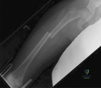
Candidate: I would measure the narrowest part of the medullary canal (the isthmus). The combined diameter of the two nails should be approximately 80% of the isthmus width. I would pre-bend the nails into a smooth arc, with the apex at the fracture site, to provide three-point fixation.
Candidates often forget the specific math (80% rule/40% per nail) or fail to mention that the apex of the bow must be precisely at the fracture site. Some candidates fail to specify that both nails must be of the same diameter, which is critical for balanced stability.
The candidate should state: "I measure the narrowest medullary isthmus; the sum of the two nails should fill 80% of the canal (each nail diameter = 40% of isthmus width). Biomechanically, the nails must be of identical diameter to prevent the stiffer nail from causing malalignment. I contour a continuous bow with the apex at the fracture site, ensuring the curve height is approximately 3x the canal diameter. This creates symmetric three-point fixation and generates the necessary outward radial force to achieve secondary bone healing via controlled micromotion."
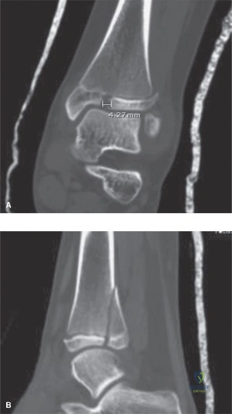
During your distal femoral entry, you are concerned about potential complications related to the hardware. What are the key technical steps to perform the distal portal creation and closure to minimize the risk of "hardware prominence" and long-term knee morbidity?
Candidate: I would ensure my entry point is at least 2-3 cm proximal to the distal femoral physis. During closure, I would cut the nails to 1-1.5 cm from the cortex, ensure they are buried beneath the fascia, and repair the IT band over the lateral hardware to prevent snapping syndrome.
Failure to explicitly mention the protection of the physis (iatrogenic growth arrest) or forgetting to close the fascia/IT band over the nails. Some candidates fail to mention that the nails must sit flush against the metaphyseal flare to avoid bursitis.
The candidate should structure the response: 1) Physeal protection: Entry points 2.5-3.0cm proximal to the physis, using fluoroscopy. 2) Hardware seating: Nails cut to 1-1.5cm from the cortex and tapped flush to the metaphyseal flare. 3) Soft tissue management: Explicitly burying the nail ends beneath the deep fascia and performing a meticulous repair of the IT band over the lateral nail to avoid painful snapping/bursitis, followed by layered closure of the vastus medialis.
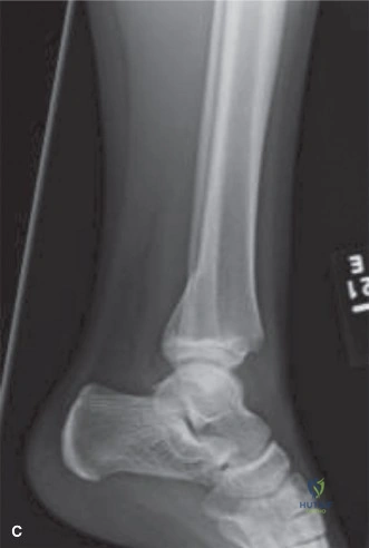
The patient is now 3 months post-operative. Radiographs show good bridging callus. The family asks about the "corkscrew effect" and when they can return to contact sports. How do you address these concerns?
Candidate: The "corkscrew effect" occurs if the nails cross at the fracture site rather than in the metaphyses, leading to rotation. Regarding sports, I would wait for complete cortical remodeling, typically around 3-4 months, before clearing for contact activities.
Ignoring the clinical timeline or allowing early return to contact sports before radiographic evidence of full remodeling. Failing to define that the corkscrew effect is a rotational stability issue caused by improper crossing points.
The candidate should explain: 1) Corkscrew mechanism: It is a failure of rotational stability caused by the nails crossing at the fracture site. Proper technique requires them to be divergent or cross only in the metaphyseal regions. 2) Return to activity: Return to non-contact activity is permitted once bridging callus is seen (6 weeks). However, return to contact sports like football requires radiographic evidence of cortical continuity and complete remodeling, which is typically 3-4 months. Hardware removal is considered at 6-12 months.
