8 Pediatric Cases: Critical Decisions on Exits from the Physis
Key Takeaway
This topic focuses on 8 Pediatric Cases: Critical Decisions on Exits from the Physis, For a 7-year-old girl with a length-stable femur fracture, reduction and flexible nail fixation is the optimal treatment. The most common complication parents should be informed about is pain/irritation at insertion sites. While flexible nails are carefully positioned, understanding the implications of how hardware *exits from the physis* is critical in pediatric orthopedics to prevent potential growth abnormalities.
You are in the trauma bay. A 7-year-old child presents with a high-energy femoral shaft fracture following a football tackle. Initial assessment confirms hemodynamic stability. You are presented with the initial AP/Lateral radiographs. How do you approach the management of this patient, and what is your reasoning for the choice of fixation?
Candidate: I would perform a full ATLS survey to rule out polytrauma. Since this is an isolated fracture in a 35kg, 7-year-old child, I would avoid a spica cast due to her weight and the risk of malunion. I would opt for Flexible Intramedullary Nailing (FIN) because she is in the appropriate weight/age bracket and the fracture is length-stable.
Failing to mention the specific weight-based contraindications for spica casting or adult rigid nailing. Candidates often forget to mention the neurovascular exam or the assessment of the ipsilateral hip and knee, assuming the injury is isolated without stating why.
Start with a systematic approach: "My priority is a primary survey to rule out life-threatening injury. Given she is 7 years old and 35kg, my management algorithm is guided by the AAOS guidelines." State clearly: "Spica casting is contraindicated due to the risk of shortening and nursing burden. Rigid intramedullary nailing is contraindicated due to the risk of AVN of the femoral head and damage to the trochanteric apophysis. Therefore, I propose Titanium Elastic Nails (TENs). I would confirm the 'personality' of the fracture using in-traction films to ensure it is length-stable and would template for the nails to occupy 80% of the isthmus diameter."
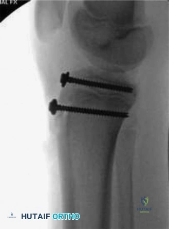
You have decided to proceed with Titanium Elastic Nailing (TENs). Explain the biomechanical principles and the critical technical steps to avoid common complications during this procedure.
Candidate: The principle is three-point fixation. I would use two nails of the same diameter, pre-contoured, to create symmetric bending moments. I must ensure they cross proximal to the fracture site and anchor in the proximal metaphysis without crossing the physis.
Ignoring the danger of the entry point relative to the distal physis. Candidates often forget that the entry must be 2.5–3cm proximal to the physis to prevent iatrogenic arrest, or they use mismatched nail sizes, which leads to hardware failure or loss of reduction.
Discuss the "Symmetric Bending Moment": "The nails act as internal splints through three-point fixation. To achieve this, the nails must be of equal diameter, and the apex of the pre-bend should be at the fracture site. Crucially, I will insert them through a metaphyseal entry point 2.5-3cm from the physis to avoid physeal injury. They must cross proximal to the fracture, and I will ensure the nails diverge proximally—one into the greater trochanter and one into the femoral neck—to provide rotational stability. Finally, I will cut the nails to leave 1-2cm protruding to allow for future removal."
Following the surgery, the parents ask about the potential for future limb length discrepancy and the timing of hardware removal. How do you counsel them?
Candidate: I would explain that overgrowth is expected, usually 1-2cm, due to the fracture healing process. I would suggest removing the hardware once the fracture is fully consolidated, usually around 6 months, or if they become symptomatic.
Dismissing the concern as "normal." Failure to quantify the overgrowth risk or failing to mention that the nails *must* be removed because they are not permanent implants in a growing child.
Structure the answer into "Overgrowth" and "Hardware Management": "I would counsel the parents that pediatric femur fractures typically undergo a period of post-traumatic overgrowth, averaging 1-2cm, which is a known phenomenon due to hyperemia at the fracture site stimulating the physes. Regarding the hardware, titanium elastic nails are not permanent implants. They serve as a temporary scaffold and are associated with soft tissue irritation. I schedule elective hardware removal once radiographic union is confirmed, typically at 6–9 months, or earlier if the child experiences bursitis or local pain."
