Pediatric Fracture Cases: Clinical Presentation, Exam, & Imaging Insights
Key Takeaway
Pediatric fractures, like distal radius, supracondylar humerus, and Salter-Harris type II, are assessed through detailed patient history, mechanism of injury, and thorough clinical examination. This includes vital neurovascular checks, inspection for deformity, palpation for tenderness, and range of motion. Orthogonal radiographs are crucial for diagnosis and classification.
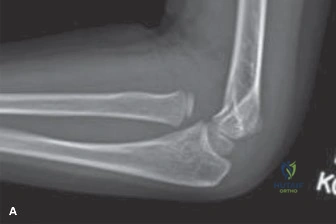
A 6-year-old female presents after falling from monkey bars, landing on an outstretched hand. She is in severe pain with an elbow deformity. Initial assessment reveals an absent radial pulse with a capillary refill time of 3 seconds. Describe your immediate management and the surgical priority.
Candidate: I would immediately document the neurovascular status and ensure the arm is splinted. Since the radial pulse is absent, this is a "pulseless supracondylar fracture." I would take her to the operating theatre for urgent closed reduction and percutaneous pinning (CRPP). If the pulse doesn't return after reduction, I would consider open exploration of the brachial artery.
Failing to emphasize the urgency of the "pink, pulseless" hand vs. the "white, pulseless" hand. A borderline candidate often fails to mention that if the hand is perfused (pink), reduction is the priority, not vascular exploration. Also, failing to state that closed reduction must be performed under general anesthesia is a missed opportunity to show clinical maturity.
Structured approach: 1. Confirm neurovascular status and document findings. 2. Recognize this as a Gartland Type III fracture. 3. Manage the 'pulseless' hand: If the hand is warm and pink, the priority is urgent closed reduction and pinning to relieve kinking of the brachial artery. If the hand remains pulseless after successful reduction, then open exploration is mandatory. 4. Pinning configuration: Use two lateral pins or cross-pins, emphasizing avoiding the ulnar nerve (avoiding medial pin insertion unless absolutely necessary or under direct visualization).
A 12-year-old male presents with this ankle injury following a soccer tackle. What is the classification, and what is your specific concern regarding his future growth?
Candidate: This is a Salter-Harris Type II fracture of the distal tibia. The concern is growth arrest or physeal bar formation leading to angular deformity or leg length discrepancy, so I would aim for anatomical reduction.
Ignoring the "Thurston-Holland" fragment's role. Candidates often fail to mention that they will counsel the parents about long-term follow-up (up to skeletal maturity) to monitor for physeal closure—this is the most important "consultant-level" detail.
Identify: Salter-Harris Type II injury with the Thurston-Holland fragment. Management: Closed reduction under sedation/GA, aiming for anatomical reduction to reduce the risk of premature physeal closure. Crucially, emphasize the need for clinical and radiographic follow-up for 6-12 months (or until skeletal maturity) because even with good reduction, growth arrest remains a possibility.
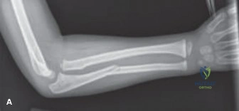
A 10-year-old girl has this forearm injury. The deformity is significant. When would you move from attempting a closed reduction in the ED to operative intervention?
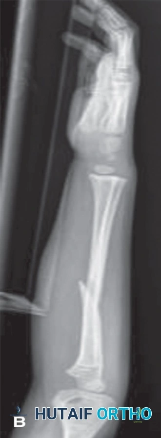
Candidate: I would attempt a closed reduction. I would move to surgery if the reduction is unstable in the cast, if there is unacceptable angulation on repeat X-ray, or if there is neurovascular compromise.
Giving vague definitions of "unacceptable." Candidates need to articulate specific parameters: angulation in the plane of motion, loss of reduction on follow-up radiographs, or persistent neurovascular deficits post-reduction.
State: "My decision is guided by the loss of reduction criteria." Specifically: 1. Irreducible fracture. 2. Failure to maintain alignment in the cast (measured at 1-week follow-up). 3. Unacceptable angulation (generally >15-20 degrees depending on proximity to physis and age). 4. Persistent neurovascular compromise. Mention that for a 10-year-old, the remodeling potential is decreasing, necessitating more precise alignment than a younger child.
