Complex Pediatric Distal Tibial Physeal Fracture: Risks of Single-Sport Specialization

Key Takeaway
Complex pediatric distal tibial physeal fractures are diagnosed via clinical exam, plain X-rays, and essential CT scans. CT reveals precise fracture morphology, displacement, and articular involvement. This detailed imaging is critical for optimal treatment, especially for injuries linked to single-sport specialization in young athletes.
A 14-year-old male presents with an acute ankle injury after a soccer injury. He has a history of vague distal tibia pain. Based on this presentation and the provided radiograph, what is your primary diagnosis, and what anatomical features predispose this age group to this specific injury?
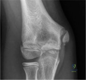
Candidate: This looks like a triplane fracture of the distal tibia. This occurs in adolescents because the physis is closing. Specifically, it closes from central to medial, leaving the anterolateral aspect open and vulnerable to rotational forces during sports.
Failing to mention the "Kump bump" or the specific sequence of physeal closure (central -> medial -> lateral). Also failing to connect the patient's history of "shin splints" (stress reaction) to the mechanism of fracture, and simply describing the radiograph without suggesting further imaging.
Acknowledge the diagnosis as a Triplane Fracture. Explain the anatomy: the distal tibial physis closes asynchronously. In Tanner Stage IV, the central/medial segments are fused, while the anterolateral aspect remains cartilaginous. The external rotation force causes the anterior inferior tibiofibular ligament (AITFL) to pull off the anterolateral epiphysis. Furthermore, identify that the patient's prodromal "shin splint" pain indicates a pre-existing metaphyseal stress reaction, which lowers the threshold for catastrophic failure under torsional load.
You decide to move forward with operative management. What imaging is mandatory, and why is plain radiography often insufficient in this injury?
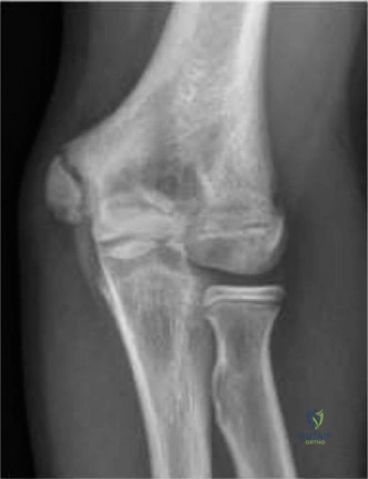
Candidate: I would order a CT scan of the ankle. Plain films don't show the true displacement, and I need to see the coronal and sagittal planes to plan my reduction and screw placement.
Stating that the CT scan is "for confirmation." A strong candidate must specify that plain films frequently underestimate articular displacement—often by more than 2mm—and fail to show the full extent of the complex three-plane involvement (sagittal, axial, and coronal).
A CT scan with sagittal, coronal, and 3D reconstruction is mandatory. Plain radiographs miss the true articular step-off in up to 40% of cases. The CT is necessary to map the three-plane injury: the sagittal epiphyseal, axial physeal, and coronal metaphyseal fracture lines. This allows for precise surgical planning regarding fragment size, articular congruity, and the trajectory of fixation to avoid the physis where possible.
Describe your surgical approach and the specific steps taken to ensure an anatomic reduction. What is the most common barrier to a successful closed reduction?
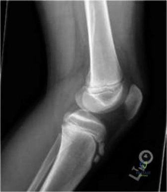
Candidate: I would use an anterolateral approach. I need to clear the fracture hematoma and any interposed periosteum, which is usually the reason closed reduction fails. Then I reduce the joint surface and fix it with screws.
Ignoring the superficial peroneal nerve branches during the approach, or failing to emphasize the "hook test" for syndesmotic integrity. Also failing to mention the use of washers for the screw heads to prevent subsidence into the soft pediatric bone.
The approach is an anterolateral incision, protecting branches of the superficial peroneal nerve. The most common barrier to closed reduction is interposed periosteum, which must be debrided. Anatomic reduction is verified visually and via fluoroscopy. Fixation involves provisional K-wires followed by cannulated, partially threaded screws (4.0/4.5mm) with washers to prevent cortical sinking. Epiphyseal screws should be parallel to the joint, and the surgeon must confirm syndesmotic stability—if the anterior inferior tibiofibular ligament is intact but the fibula is fractured, syndesmotic fixation may be necessary.
The surgery went well, but the parents are concerned about future growth. What is the long-term risk for this patient, and how would you counsel the family regarding his return to competitive sports?
Candidate: The main risk is physeal arrest and growth discrepancy, but since he is Tanner Stage IV, the growth remaining is minimal. I would tell him to stop specializing in just one sport to avoid another overuse injury.
Giving vague advice on returning to sports without mentioning "periodization" or sport-specific functional testing. Also failing to mention the risk of post-traumatic osteoarthritis due to any residual articular incongruity.
Long-term risks include premature physeal closure leading to angular deformity or leg length discrepancy, and post-traumatic OA. Given he is Tanner Stage IV, the growth risk is lower, but still present. Regarding sports: emphasize periodization. I would require 3 months of off-season or cross-training annually. Return to play is conditional on full, painless ROM, symmetrical strength, and functional testing, typically at 3-5 months post-op. This is a critical opportunity to educate the family on the dangers of youth sports specialization.
