Pediatric Buckle Fracture: When is a Velcro Wrist Brace Best?
Key Takeaway
Your ultimate guide to Pediatric Buckle Fracture: When is a Velcro Wrist Brace Best? starts here. For a distal radius buckle fracture, treatment options include a short-arm cast or a prefabricated velcro wrist brace. Both effectively immobilize the fracture, which is at low risk of displacement. A velcro wrist brace often provides higher patient satisfaction and convenience, though it may cause more initial pain than a cast. Families can be offered either choice for support during healing.
An 8-year-old child presents to the ED following a FOOSH injury. They are holding their wrist, which shows minimal swelling. There is no obvious deformity. You are presented with the following radiograph. Describe the findings and your initial management plan.
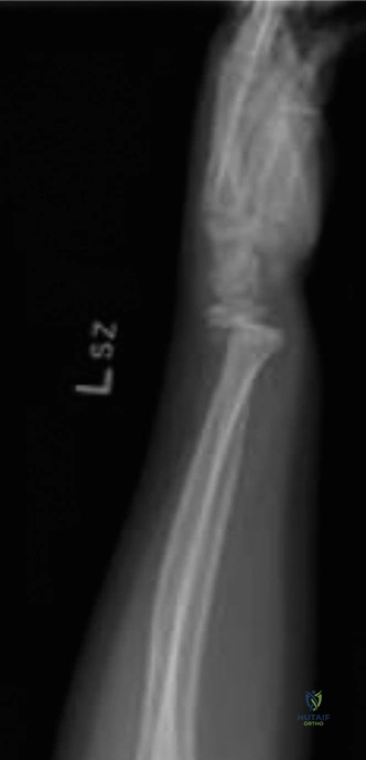
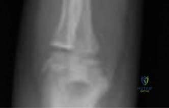
Candidate: The radiograph shows a buckle or torus fracture of the distal radial metaphysis. There is a characteristic cortical buckling or wrinkling on the dorsal aspect, with the other cortex appearing intact. This is a stable injury. I would manage this non-operatively with a Velcro wrist brace for 3 to 4 weeks and provide parental education regarding pain management and activity modification.
Failing to explicitly state the stability of the fracture or suggesting routine follow-up radiographs. Some candidates over-complicate the answer by suggesting a formal plaster cast, which is considered over-treatment in modern evidence-based practice for this specific, stable injury.
Identify the injury as a stable distal radial metaphyseal buckle (torus) fracture. Emphasize the biomechanics (axial load, compression of the cancellous metaphysis with an intact periosteum). Support non-operative management with a Velcro brace based on current high-level evidence, noting that routine follow-up X-rays are unnecessary. Mention the exclusion of "red flags" (complete cortical breach, physeal involvement, or neurovascular deficits).
The parent asks why you are not applying a full plaster cast, as their neighbor's child had one for a similar-looking injury. How do you address this parental concern while maintaining clinical rigor?
Candidate: I would explain that their child has an inherently stable 'buckle' fracture. I would cite evidence demonstrating that Velcro braces are equivalent to casting in terms of healing and long-term function, while significantly improving patient comfort, hygiene, and compliance. I would assure them that this approach prevents the risks associated with rigid immobilization, such as skin issues or unnecessary stiffness.
Dismissing the parent's concern or simply stating "the cast is unnecessary." A high-scoring candidate demonstrates communication skills by validating the parent's anxiety while pivoting to the evidence-based benefits of the brace.
Structure the response: 1. Validate the parent's concern. 2. Define the stability of the injury. 3. Summarize the literature (e.g., mention RCTs/meta-analyses showing equal outcomes). 4. List the patient-centered benefits (hygiene, comfort, cost-effectiveness, reduced hospital visits).
When does a distal radius injury in a child cross the line from a simple, brace-managed buckle fracture to an injury requiring more aggressive, potentially operative, management?
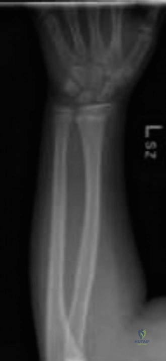
Candidate: An injury ceases to be a simple buckle fracture if there is a complete cortical breach (e.g., greenstick or complete fracture), significant angulation (>15-20 degrees), rotational deformity, or if it involves the physis (Salter-Harris type III, IV, or V). Furthermore, any associated neurovascular deficit or open injury mandates urgent surgical consideration.
Failing to mention the specific threshold for angulation or forgetting to classify the physeal involvement. Also, failing to identify "impending neurovascular compromise" or "open injury" as absolute markers for higher-level intervention.
Provide a structured classification: 1. Anatomical/Fracture Stability: Complete vs. Incomplete cortex, angulation (>15-20°), and rotatory alignment. 2. Physeal Involvement: Differentiate between simple SH I/II (casting) and complex SH III/IV (surgical). 3. Clinical Red Flags: Neurovascular compromise, open wounds, or compartment syndrome. 4. Patient Factors: Non-compliance necessitating rigid fixation.
