Middle-Third Clavicle Fractures: An Exhaustive Review of Anatomy, Pathophysiology, and Management
Key Takeaway
Middle-third clavicle fractures are frequent traumas exhibiting specific displacement patterns. While non-operative historically, operative fixation is now favored for significantly displaced or comminuted cases to reduce symptomatic malunion and nonunion. Thorough neurovascular assessment and biomechanical understanding are crucial for optimizing patient outcomes.
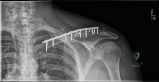
A 28-year-old male cyclist presents to the emergency department following a high-speed fall onto his right shoulder. He complains of severe pain and inability to move the arm. Examination reveals a visible deformity in the mid-portion of the clavicle, with tenting of the skin. Neurovascular examination is intact. The following radiograph is obtained.
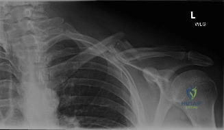
How would you classify this fracture, and what are the specific clinical factors that would influence your management decision?
Candidate: I would classify this as a Robinson Type 2B2 midshaft clavicle fracture because it is displaced and comminuted. Management depends on the patient's activity level and the degree of displacement/shortening. Given he is a young, active male with clear displacement, I would likely recommend operative fixation, especially since there is tenting of the skin.
Failing to mention the "shortening" measurement. Candidates often focus purely on the displacement or the "type" without quantifying the shortening (which is a critical independent predictor of nonunion). Additionally, failing to address the "tenting" as a potential emergency or indication for urgent surgical decompression is a significant oversight.
Structure the answer by: 1) Classification: Confirming Robinson Type 2B2. 2) Decision Matrix: Specifically cite the threshold of >2cm of shortening and 100% displacement as markers for increased nonunion risk. 3) Clinical Urgency: Note that the skin tenting represents a "threatened soft tissue envelope," which may necessitate more urgent intervention to prevent skin breakdown, regardless of the Robinson score. 4) Evidence: Mention that the COTS (Canadian Orthopaedic Trauma Society) study supports operative fixation for displaced midshaft fractures to improve functional outcomes and reduce nonunion compared to conservative management.
You have decided to proceed with Open Reduction and Internal Fixation (ORIF). During your dissection, you identify several cutaneous branches crossing the field. As you approach the bone, describe how you mitigate the risk of injury to the underlying neurovascular structures.
Candidate: I would identify and protect the supraclavicular nerves early by retracting them with vessel loops. To protect the subclavian vessels, I would place a periosteal elevator or a blunt retractor deep to the clavicle during all drilling and screw insertion steps. I also prefer using oscillating drill bits and depth gauges to avoid plunging.
Candidates often forget to mention the management of the supraclavicular nerves if they are in the way. Simply saying "I'll be careful" is insufficient. A poor answer also neglects the importance of the subclavius muscle as a natural anatomical buffer, which the surgeon should try to maintain as much as possible.
A high-scoring answer details the "Layered Safety Strategy": 1) Nerve Preservation: Systematic identification and retraction; if transection is unavoidable, bury the stump in local muscle to mitigate neuroma. 2) Mechanical Protection: Always utilize a sub-periosteal blunt retractor between the clavicle and the chest wall. 3) Technical Precision: Use of oscillating drill attachments, drill stops, and avoiding "over-drilling" through the posterior cortex. 4) Vascular Awareness: Acknowledge that the subclavian vessels are at their closest point in the midshaft, requiring the surgeon to avoid placing screws in a deep, postero-inferior trajectory.
