Anterior Glenohumeral Dislocation: Comprehensive Guide to Epidemiology, Anatomy, Biomechanics & Management
Key Takeaway
Anterior glenohumeral dislocation is when the humeral head displaces anteriorly from the glenoid, representing 95-97% of all shoulder dislocations. Often caused by an abducted, externally rotated arm trauma, it involves injury to static stabilizers like the Inferior Glenohumeral Ligament complex (e.g., Bankart lesion) and potential axillary nerve damage.
A 22-year-old rugby player presents to the ED after a tackle, holding his arm in slight abduction and external rotation. He is in significant pain. Describe your immediate management priorities in the emergency department.
Candidate: I would perform a primary survey, then a neurovascular assessment, followed by radiographs (AP and Scapular Y views). After obtaining informed consent, I would provide adequate analgesia and sedation to perform a closed reduction. Finally, I would re-examine his neurovascular status and obtain post-reduction films.
Failing to emphasize the pre-reduction neurovascular examination. Many candidates forget to document axillary nerve function (sensation over the regimental badge area) and distal pulses before the manipulation, which is medico-legally critical.
Structure the answer: 1. Assessment (History, examination, meticulous pre-reduction neurovascular assessment). 2. Imaging (Standard trauma series: AP and Y-scapular; comment on why these are needed). 3. Procedural Safety (Informed consent, appropriate analgesia/sedation). 4. Execution (Gentle, controlled reduction technique). 5. Post-reduction (Neurovascular check, confirmation of reduction via X-ray, and immobilization).
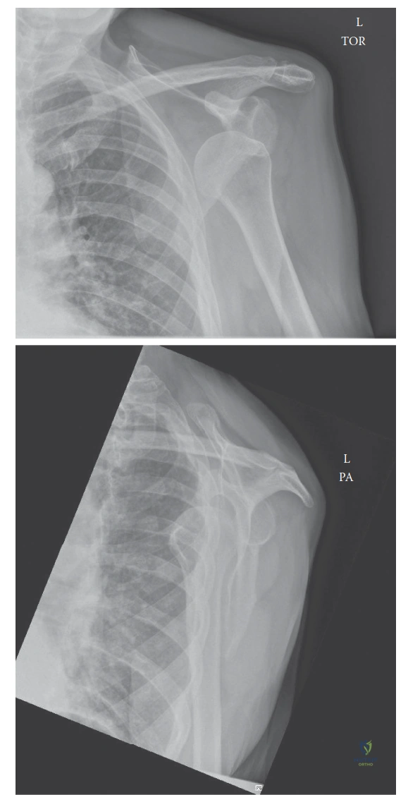
The patient has been successfully reduced. You are presented with the post-reduction radiograph below. What findings do you note, and how do they influence your counseling regarding future stability?
Candidate: The radiograph shows a congruent joint. I am looking for a Hill-Sachs lesion on the humeral head or a bony Bankart on the glenoid. In a young athlete, this first episode puts him at high risk of recurrence, so I would discuss the necessity of early physiotherapy or potential early surgical stabilization if risk factors like glenoid bone loss are present.
Providing a vague management plan. Examiners expect you to quantify the "risk of recurrence" based on age and activity. Failing to mention CT imaging for bony defects in an athlete is a significant oversight.
Identify findings: "I see a congruent reduction with a visible Hill-Sachs lesion on the humeral head." Then, integrate the clinical context: "For a 22-year-old contact athlete, the risk of recurrence is high (up to 80-90%). I would counsel him on the 'three pillars' of management: activity modification, comprehensive rehabilitation, and the potential role of early arthroscopic stabilization to prevent secondary bone loss and soft tissue damage."
What are the absolute contraindications for a blind closed reduction maneuver in the ED?
Candidate: Absolute contraindications include signs of neurovascular compromise such as absent distal pulses or ischemia, as well as an open dislocation. Also, if there is a suspected fracture of the surgical neck of the humerus, I would proceed with extreme caution or request senior input.
Missing "irreducible dislocation" caused by soft tissue interposition. Candidates often focus only on bony contraindications while ignoring the clinical reality that some shoulders are mechanically locked.
Categorize for clarity: 1. Neurovascular (Absent pulses, acute ischemia requiring vascular surgery). 2. Soft Tissue/Mechanical (Suspected irreducible dislocation due to interposition of the biceps, labrum, or subscapularis). 3. Bony (Complex fracture-dislocations where manipulation might convert a stable fracture to an unstable one, or open dislocations).
